


Figure 4. Co-localization and heterodimerization of TLR11 and TLR12.
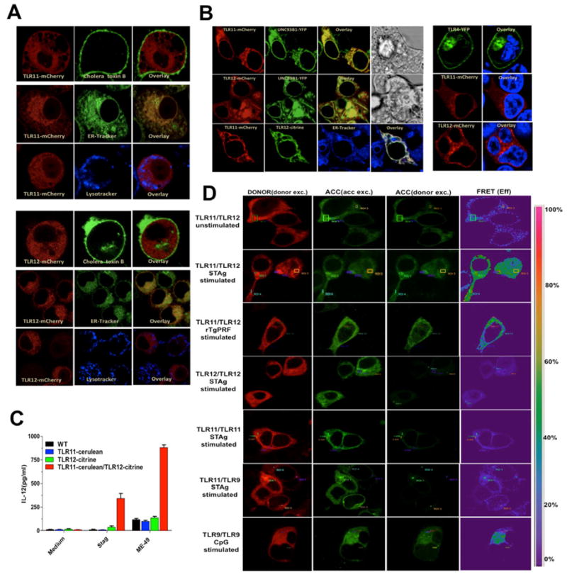
(A) Immortalized macrophages were stably transfected with TLR11- or TLR12-mcherry and imaged post-staining with Cholera toxin B subunit FITC conjugate, LysoTracker White-Blue or ER-Tracker White-Blue. (B) HEK 293T cells transfected with TLR11-mCherry or TLR12-mcherry with UNC93B1-YFP or TLR11-mcherry and TLR12-citrine (left panel) or with TLR4-YFP and stained with Hoechst 33342 as nuclear marker (right panel). (C) Immortalized macrophages stably expressing either TLR11, TLR12 or both were stimulated in vitro with STAg (10 μg/ml) or exposed to live tachyzoites (MOI 3:1) of the ME49 strain and levels of IL-12p40 measured in the supernatants at 24 h post-stimulation. Data are represented as mean ± SD of three experiments. (D) HEK 293T cells were transfected with the plasmids encoding the proteins indicated in the figure; 48 h after transfection, cells were left unstimulated or were stimulated with STAg (10 μg/ml), rTgPRF (100 ng/ml) or CpG 1826 (1 μM) for TLR9/TLR9. FRET between the respective proteins was calculated by measuring sensitized emission (SE) fluorescence using the FRET SE wizard on the Leica SP2 confocal laser-scanning microscope. For each plasmid combination, cerulean (represented in red) was used as donor and citrine (represented in green) as acceptor. Data are from one representative experiment of four.