


Figure 3. Association of torsinA and Derlin-1 in primary neurons.
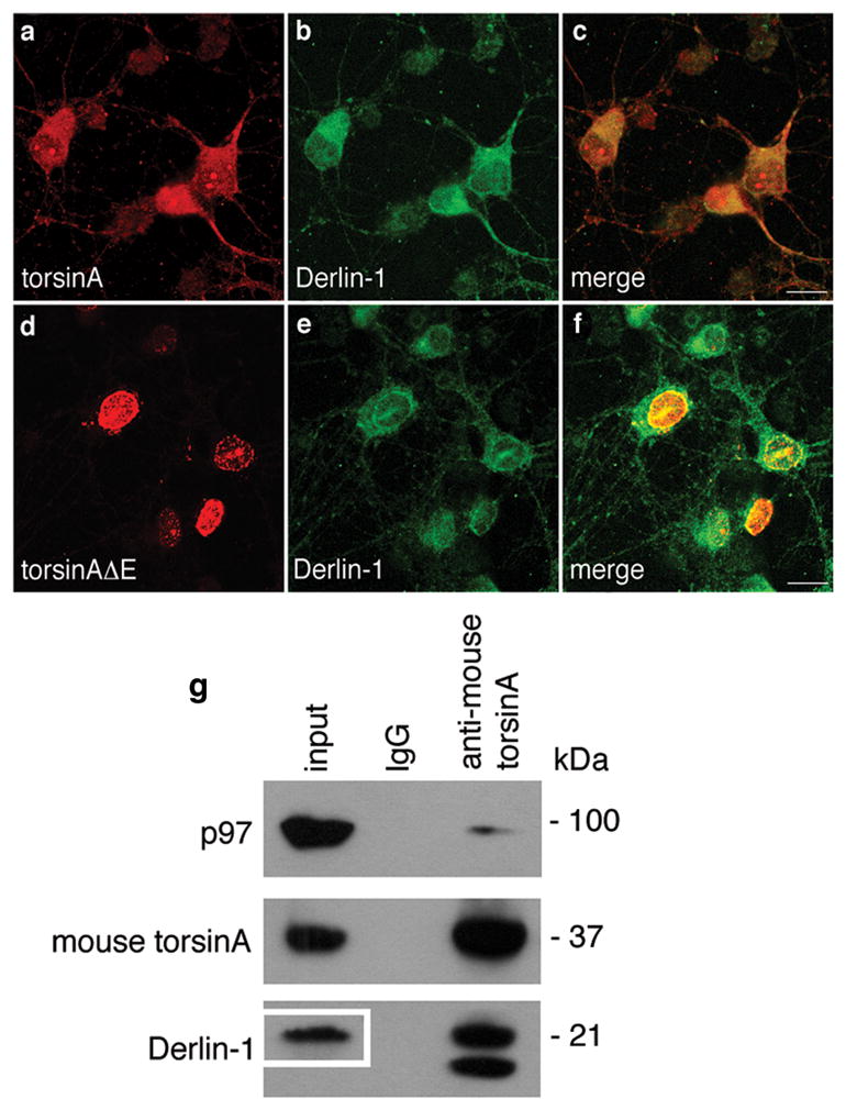
Neuronal cultures were prepared from E15 mouse embryos. (a-f) Established cultures were infected overnight with (a-c) AAV-CBA-torsinA-GFP or (d-f) AAV-CBA-torsinAΔE-GFP at an M.O.I. of 50 genome copies per cell. Seventy h later immunocytochemistry was performed for (a & d) torsinA and (b & e) Derlin-1, with merged images shown in panels (c) and (f), respectively. Representative neurons shown. Magnification bar = 10 μm. (g) Neurons expressing only endogenous proteins were harvested and lysates immunoprecipitated with IgG or antibodies to mouse torsinA. Immune precipitates were resolved by SDS-PAGE and immunoblotted with antibodies to p97, mouse torsinA and Derlin-1. In the total lysate (input) so little immunoreactive Derlin-1 observed after the 2 min exposure that the blot had to be re-exposed for 90 min to visualize this protein (see inset box in input lane). For Derlin-1 the top band has the correct MW for this protein, an additional immunoreactive lower band was seen only in torsinA immune precipitates from neuronal cultures.