


FIGURE 3.
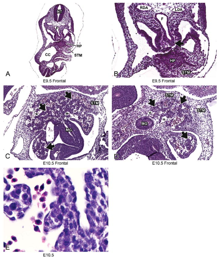
Representative images of E9.5 and E10.5 embryonic mouse liver. Low-magnification H&E-stained frontal sections at E9.5 (A, B) and E10.5 (C, D). High magnification at E10.5 (E). Histologically, liver development begins around E9.5 as a diverticulum of proliferating endodermal cells arising from the ventral surface of the caudal foregut (A, B, arrow). By E10.5 (C, D), the epithelial sheets of the liver bud have undergone elongation and branching to form hepatic cords (arrows) separated by blood-filled sinusoids (asterisks). The hepatic cords delaminate from the foregut and move into the septum transversum mesenchyme at this stage. At high magnification, clear vacuoles can be seen within the hepatoblast cytoplasm and nucleated red blood cells can be seen within the sinusoids (E). CC = coelomic cavity; F = foregut; HP= hepatic primordium; LDA = left dorsal aorta; MG = midgut; NT = neural tube; RDA = right dorsal aorta; STM = septum transversum mesenchyme.