


Figure 9.
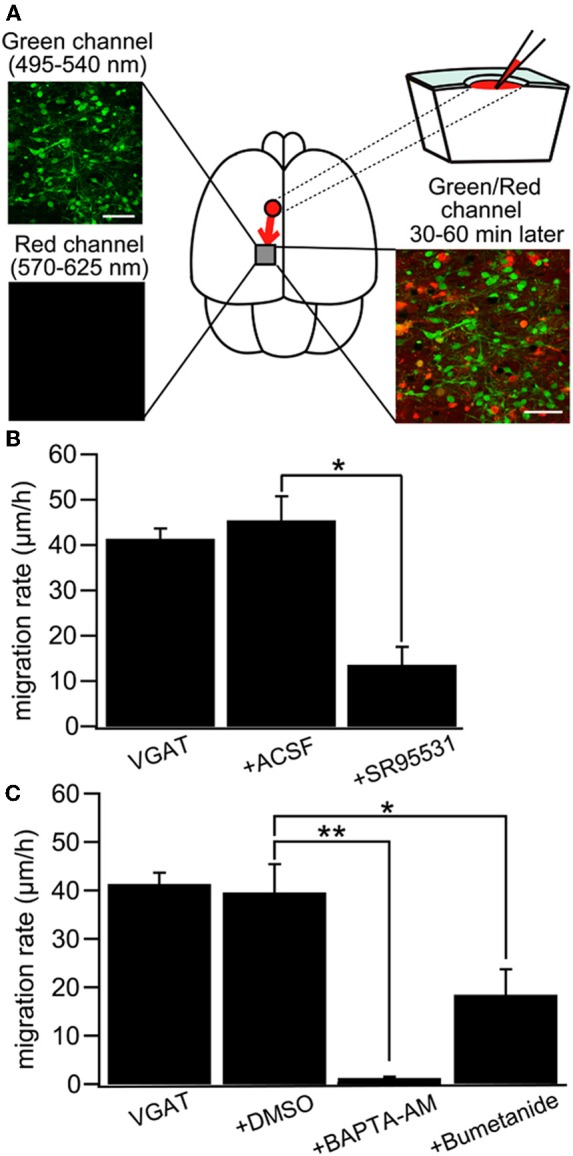
GABA regulates tangential migration in the immature cortex in vivo. (A) Upper left: Two-photon image of Venus positive cells immediately after drug application in a P0 VGAT-Venus mouse. Making a hole at 1–2 mm away from the observed area has no effect on the morphology of the cells within imaging area. Scale bar = 50 μm. Lower left: Two-photon image of sulforhodamine 101 (SR101) showing that the drug does not reach the imaging area immediately after injection. Upper right: Schematic drawing of the drug injection procedure; making a hole and injecting the drug conjugated with SR101. The drug diffuses into the imaging area after 30–60 minutes of injection. Lower right: Two-photon image of Venus-positive cells and SR101 showing that the pipette solution spread around the cells without injuring them. Scale bar = 50 μm. (B) Cell displacement during 1 h of migration for neurons in VGAT-Venus without drug application (VGAT), after injection of ACSF (+ACSF) and after injection of SR95531 (+SR95531). The selective GABAAR antagonist (SR95531, 1 mM, 1 μl) reduced the migration rate significantly (*p < 0.05) whereas ACSF (1 μl) had no effect. (C) DMSO (1 μl) had no effect on cell displacement. Chelating intracellular Ca2+ by BAPTA-AM (2 mM, 1 μl) conjugated with DMSO almost completely inhibited the migration (**p < 0.01) and blockade of NKCC by bumetanide (1 mM, 1 μl) significantly reduced the migration rate (*p < 0.05). [From Inada et al. (2011)].