


Figure 2. Morphological feature of phagocytose and autophagy in eukaryotic cells.
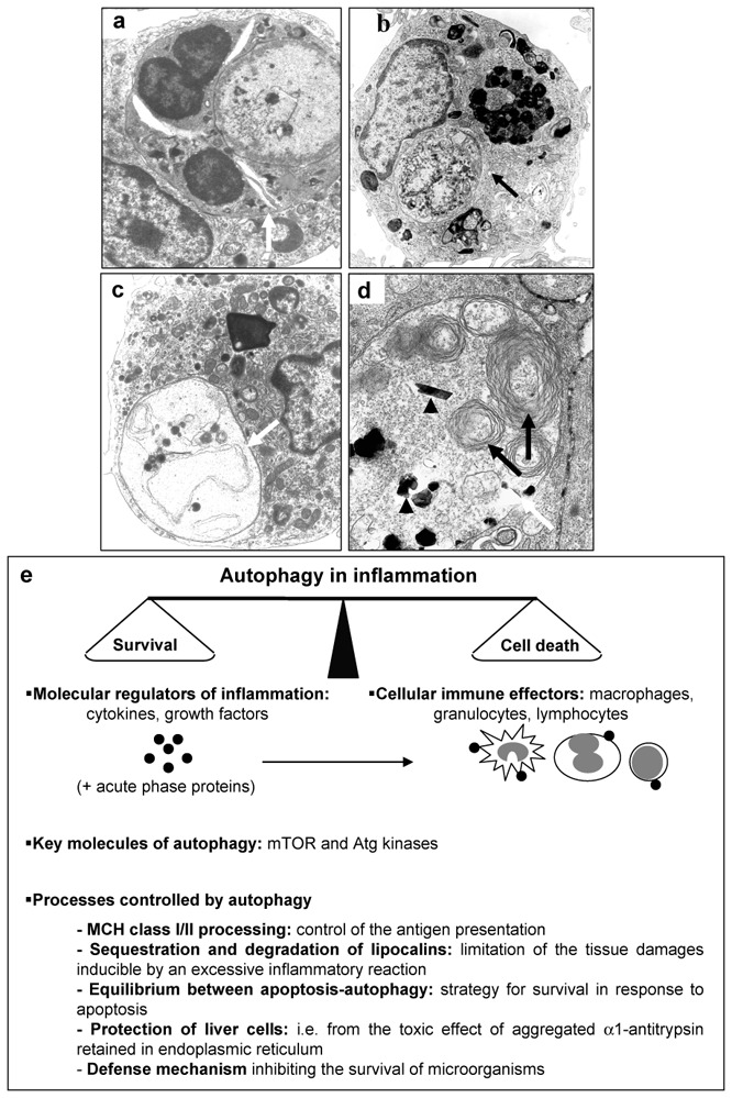
(a) Phagocytose of a polynuclear (white arrow) by a macrophage; (b, c) typical feature of autophagy (white and black arrows) into macrophages in culture; (d) macrophage in culture degrading calcium phosphate ceramic, autophagic vesicle (white arrow) (containing cell endogenous materials (black arrow) and exogenous materials (black arrow head). Original magnification: (a, c) X 7,500; (b) X 5,000; (d) X 11,000; (e) cellular/molecular protagonists involved in autophagy associated with inflammation and the main processes controlled by autophagy during inflammatory process.