



Abstract
Mechanisms governing secretion of proteins underlie the biologic activities and functions of human eosinophils, leukocytes of the innate immune system, involved in allergic, inflammatory, and immunoregulatory responses. In response to varied stimuli, eosinophils are recruited from the circulation into inflammatory foci, where they modulate immune responses through the release of granule-derived products. Transmission electron microscopy (TEM) is the only technique that can clearly identify and distinguish between different modes of cell secretion. In this review, we highlight the advances in understanding mechanisms of eosinophil secretion, based on TEM findings, that have been made over the past years and that have provided unprecedented insights into the functional capabilities of these cells.
Keywords: transmission electron microscopy, electron tomography, eosinophils, cell secretion, vesicular transport, inflammation, immune system, cytokines
Introduction
Since the pioneer works of Palade and collaborators (Jamieson & Palade, 1967; Zetterstrom, 2006), which used electron microscopy to characterize, for the first time, organelles and interactions between various compartments of secretory pathways, cell secretion studies have greatly contributed to our understanding of physiological and pathological events. In cells from the human immune system such as eosinophils, mechanisms governing secretion underlie different biologic activities and responses to allergic and inflammatory diseases (reviewed in Moqbel & Coughlin, 2006; Melo et al., 2008c).
Eosinophil responses involve secretion of distinct cationic proteins and numerous cytokines with multiple functional activities (reviewed in Gleich, 2000; Adamko et al., 2005; Hogan et al., 2008). These mediators are released in a tightly orchestrated manner to regulate the progression of an immune response. Different from other immune cells, such as most lymphocytes that must exclusively synthesize proteins prior to secretion, both cationic proteins and cytokines are additionally stored as preformed pools within eosinophil secretory granules (Spencer et al., 2009).
Eosinophils are able to release numerous proteins from their cytoplasmic granules through different secretion modes: (1) classical granule exocytosis by which granules release their entire contents following granule fusion with the plasma membrane, including compound exocytosis, which also involves intracellular granule-granule fusion before extracellular release; (2) piecemeal degranulation, a process for secretion from intracellular granules mediated by transport vesicles; and (3) cytolysis, which involves the extracellular deposition of intact granules upon lysis of the cell (reviewed in Erjefalt & Persson, 2000; Moqbel & Coughlin, 2006).
Developments in light microscopy, in particular in confocal microscopy, have provided significant information on intracellular membrane trafficking and secretion. However, transmission electron microscopy (TEM) is the only technique with resolution sufficient to clearly identify and distinguish between different modes of cell secretion. We have been using TEM to understand secretion mechanisms in human eosinophils during different situations (Melo et al., 2005a, 2005b, 2005c, 2009; Spencer et al., 2006). Conventional TEM in conjunction with immunonanogold EM and electron tomography has helped to elucidate the structural organization of eosinophil secretory organelles and to identify a distinct vesicular system, including large tubular carriers, actively involved in intracellular trafficking and secretion of specific proteins from secretory granules (reviewed in Melo et al., 2008a, 2008c). In this review, we highlight the advances in understanding mechanisms of eosinophil secretion, based on TEM findings, that have been made over the past years and that have provided unprecedented insights into the functional capabilities of these cells.
A Novel View of Eosinophil Secretory Granules: From Storage Stations to Compartmentalized Highly Active Organelles
The secretory (also termed specific, crystalline, or secondary) granules of eosinophils are notable not only for their ultrastructural morphology but also because they store a large number of preformed proteins. These cytoplasmic granules are classically known by the presence of a central crystalline core and an outer matrix surrounded by a delimiting trilaminar membrane (Fig. 1A). Because of their unique morphology, this granule population defines the eosinophil lineage in multiple species (Dvorak & Weller, 2000).
Figure 1. (Color online) Ultrastructure of activated human eosinophils.
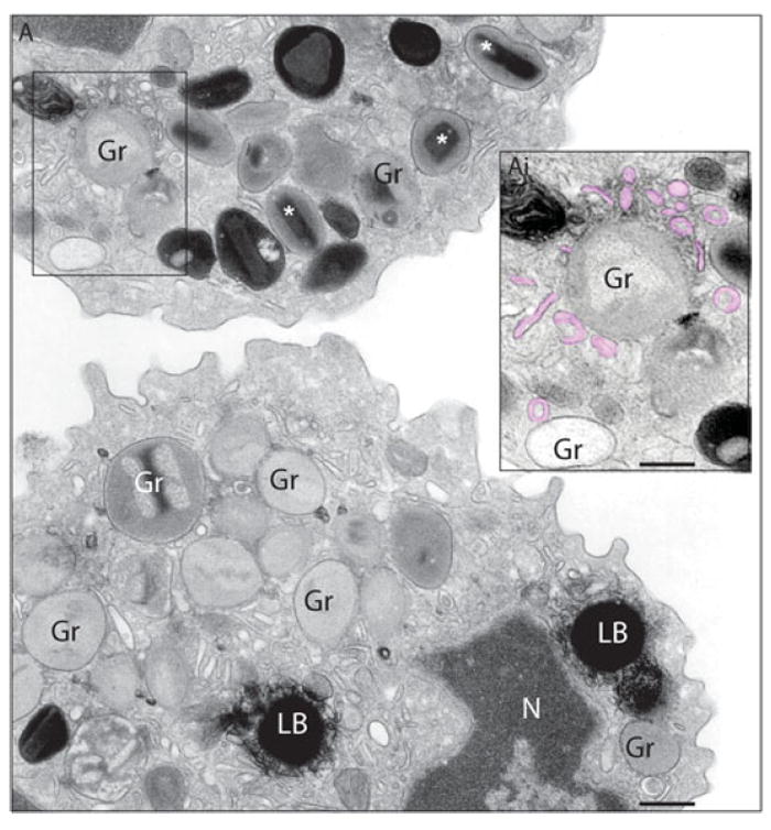
A: After stimulation with an eosinophil agonist (eotaxin), secretory granules (Gr) exhibit different degrees of emptying of their contents and show morphological diversity indicative of piecemeal degranulation. These granules show disassembled matrices and cores or residual cores or lucent areas in their cores, matrices, or both. Emptying granules are generally larger than resting granules (*), which show typical morphology with a welldefined electron-dense crystalline core. The boxed area shows many profiles of small and large vesicles (EoSVs, highlighted in pink in Ai) in contact and surrounding mobilized secretory granules. Lipid bodies (LB), inflammation-related organelles (Melo et al., 2006), are also formed in response to eotaxin stimulation. Cells were incubated with eotaxin for 1 h, immediately fixed and prepared for TEM as before (Melo et al., 2005a). N, nucleus. Bar: (A) 1.0 μm; (Ai) 600 nm.
The presence of membranes within secretory granules of eosinophils was noted only occasionally in prior years from allergic subjects (Okuda et al., 1981), Crohn’s disease patients (Dvorak et al., 1980), and in platelet activating factor (PAF)-stimulated human eosinophils (Kroegel et al., 1993). More recently, by using conventional EM to study agonist-elicited granule emptying during eosinophil secretion (piecemeal degranulation), we demonstrated that secretory granules indeed contain internal membranes (Melo et al., 2005a). Notably, as secretory granule content was mobilized in activated eosinophils, as seen after 1 h of stimulation, an extensive network of tubules and vesicles was revealed within emptying granules, mainly in the matrix area (Fig. 2). The internal tubular network was imaged as an elaborate system of convoluted tubules or individual tubular profiles at times, associated with small, round, or sac-like vesicle profiles. Quantitative EM analyses demonstrated that, in parallel with a significant increase of emptying granule numbers, there were significant increases in numbers of granules showing internal membrane domains in response to classical eosinophil agonists: CCL11 (eotaxin), CCL5 (RANTES), and PAF. Moreover, membranous subcompartments, frequently seen delimiting electron dense granule products, were prominent in granules with mobilized contents, and/or were in contact with the inner surface of the granule membrane (Melo et al., 2005a).
Figure 2. (Color online) Secretory granules exhibit internal membranous domains in human eosinophils.

A: High magnification of an emptying granule (Gr) with internal membranous tubules (highlighted in blue in Ai) seen in conjunction with mobilized core. EoSVs around the granule are indicated by arrows. A budding vesicle is indicated (arrowhead). Cells were stimulated with eotaxin as before (Melo et al., 2005a) and processed for TEM. Bar, 500 nm. Reprinted from Melo et al. (2005a) with permission.
The presence within granules of membrane domains was confirmed by immunonanogold labeling for CD63 (Fig. 3A), a tetraspanin membrane protein (Melo et al., 2005a). CD63 was localized to the granule limiting membranes, as well as to the granule matrices (Fig. 3A) and tubular vesicles (Fig. 3Ai,Aii) (Melo et al., 2005a).
Figure 3. Secretory granules are highly labeled for CD63 and show membranes organized as a tubular network.
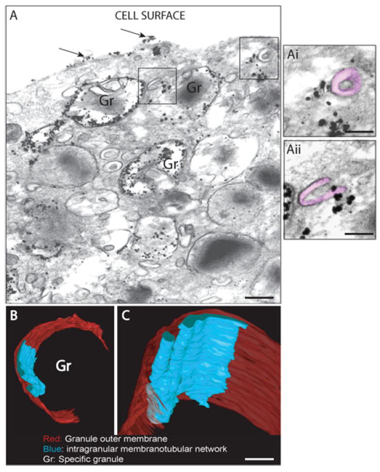
A: Immunonanogold electron microscopy revealed pools of CD63 within specific granules undergoing depletion of their contents. The boxed areas show different profiles of tubular vesicles (EoSVs highlighted in pink in higher magnification in Ai and Aii) exhibiting membrane-associated labeling for CD63. Labeling for this tetraspanin is also observed at cell surface (arrows). B, C: Show 3D models generated from serial tomographic slices (~4 nm thick) obtained from a stimulated eosinophil secretory granule analyzed by automated electron tomography (Melo et al., 2005a). The outer granule membrane is partially traced in red and intragranular vesiculotubular membranes in blue. Bar: (A) 600 nm; (Ai, Aii) 300 nm; (B) 400 nm; (C) 150 nm. Panels B and C were reprinted from Melo et al. (2005a) with permission.
To obtain greater insights into the structural mechanisms related to mobilization of intragranular products, eosinophils were studied by automated electron tomography (Melo et al., 2005a). While conventional TEM studies are usually performed on ~80-nm-thick sections, the tomographic slices are only ~4 nm thick and offer a significant advantage over typical serial “thin sections” for tracking cell structures in three dimensions (3D). Tomographic reconstructions revealed that mobilized eosinophil granule contents are rearranged within intragranular vesiculotubular compartments. Intragranular membranous subcompartments were imaged in 3D models as an aggregate of flattened tubular networks and tubules, with interconnections in some planes (Fig. 3B,C). Structural connections were revealed between the intragranular membranous network and the granule limiting membrane. This may represent membrane sites involved in granule product translocation. Intragranular structures appearing as round profiles in single routine 80-nm sections of eosinophils were revealed by the 3D models to result from cross sections of intragranular tubules rather than vesicles (Melo et al., 2005a).
Altogether, these findings suggest that proteins can be sorted within granule subcompartments before reaching the outer granule membrane in order to be delivered to the cell surface. Structural connections between the intragranular membranous network and granule limiting membrane, as visualized by electron tomography, may further contribute to the mobilization of granule proteins for their release into vesicles derived from granule surfaces. Thus, EM studies provided strong evidence for sequestration and translocation of products within eosinophil granules. A functional implication of this finding is that it adds support to the occurrence of selective release of products from eosinophils, as documented previously by us (Bandeira-Melo et al., 2001, 2003) and other groups (Lacy et al., 1999). It is now well established that differential release of cytokines occurs in response to specific stimuli (Spencer et al., 2009), and the sorting of proteins into intragranular compartments makes them easier to be delivered to cell surface through a vesicular transport.
Imaging New Transport Carriers during Immune Responses
Large Transport Vesicles for Cell Secretion
In activated eosinophils, vesicle-mediated transport of proteins from secretory granules is largely described both in vitro and in vivo during different conditions. Since the early description of this secretory pathway between the eosinophil cytoplasmic granules and cell membrane, it was believed that proteins were carried out only by small round vesicles (Dvorak et al., 1991; Dvorak, 1992; Beil et al., 1993). Only recently has light been shed on other subcellular compartments and mechanisms involved in secretion of eosinophil products. EM studies have brought conclusive evidence for the participation of morphologically distinct, large membrane-bound tubular compartments, referred to as eosinophil sombrero vesicles (EoSVs), in the eosinophil secretory route (Spencer et al., 2006; Melo et al., 2008c, 2009).
EoSVs, previously reported as microgranules (reviewed in Dvorak & Weller, 2000) or cup-shaped structures (Komiyama & Spicer, 1975) in the early eosinophil literature, represent a distinct vesicle population. They present a morphology that resembles a “mexican hat” (sombrero) when seen in conventional cross-thin sections (Figs. 2, 3Ai). EoSVs have a central area of cytoplasm and a brim of circular membrane-delimited vesicle. These tubular vesicles also exhibit a “C”-shaped morphology (Fig. 3Aii). Because of this particular morphology and large size (~150–300 nm diameter), EoSVs are easily identified by TEM in the cytoplasm of human eosinophils (reviewed in Melo et al., 2008a, 2008c) (Figs. 1, 2, 3A).
Quantitative EM studies showed that EoSVs are formed upon cell activation with eosinophil agonists such as eotaxin, RANTES, and PAF, and that these large carriers were associated with granules undergoing content release (Melo et al., 2005b). Stem cell factor (SCF), a cytokine that is also able to activate and induce degranulation in eosinophils (Oliveira et al., 2002), directs formation of EoSVs in both blood and tissue human eosinophils. Moreover, in a recent EM study, we demonstrated that the total numbers of EoSVs are significantly increased within eosinophils from a patient with hypereosinophilic syndrome (HES)(Melo et al., 2009). Eosinophils from HES individuals are typically activated (Ackerman & Bochner, 2007), compared to cells from normal donors.
Another interesting finding revealed by TEM was the fact that EoSV formation can be inhibited by brefeldin-A (BFA) (Melo et al., 2005b), a potential inhibitor of vesicular transport (Nebenfuhr et al., 2002). Studies using treatment with BFA showed that not only the total number of cytoplasmic EoSVs was reduced in the eosinophil cytoplasm, but also membranes were clearly collapsed within secretory granules (Melo et al., 2005a, 2005b). This observation pointed to a possible origin for EoSVs from secretory granules. In fact, these tubular vesicles are frequently seen attached and apparently budding from specific granules in stimulated eosinophils. A quantitative EM study showed that 90% of these granule-attached EoSVs were associated with granules showing ultrastructural changes typical of piecemeal degranulation, i.e., granules with lucent areas in their cores, matrices or both, reduced electron density, disassembled matrices and cores, residual cores, or membrane empty chambers (reviewed in Melo et al., 2008b, 2008c; Melo & Weller, 2010).
We have investigated the intriguing structure of EoSVs by electron tomography (Melo et al., 2005b). The 3D perspective that electron tomography provides revealed that EoSVs represent a dynamic system with a remarkable ability to change their shape and to interact with secretory granules (Melo et al., 2005b) (Fig. 4A). These vesicles present substantial membrane surfaces and are larger (150–300 nm in diameter) and more pleiomorphic than the small, spherical vesicles (~50 nm in diameter) classically involved in intracellular transport (Melo et al., 2005b, 2008c). Along the length of EoSVs, continuous, fully connected, and cylindrical and circumferential domains were identified as well as incompletely connected and only partially circumferential curved domains (reviewed in Melo et al., 2008c) (Fig. 4D,E). These two domains explain both the “C” shaped morphology of these vesicles and the presence of elongated tubular profiles very close to typical EoSV, as frequently seen in two-dimensional cross-sectional images of eosinophils (Melo et al., 2008c) (Figs. 1, 3Aii).
Figure 4. EoSVs are formed from mobilized secretory granules in activated eosinophils.

A–C: Representative serial tomographic slices (4 nm thick) obtained from electron tomography of a mobilized granule show substantial changes associated with EoSV formation. EoSVs, the lumens of which are highlighted in pink in panels Ai–Ci are imaged as open, tubular-shaped structures. Numbers on the lower right corners of the panels indicate slice numbers through the tomographic volume. D, E: 3D models, based on electron tomographic slices, show the structural organization of EoSVs (colored in pink). D: The granule limiting membrane is highlighted in blue and intragranular membranes in green. E: The model shows the corresponding boxed area in panel Ci. F, G: Immunonanogold EM for major basic protein (F) and interleukin-4 (G) reveals labeling at EoSV lumen and membranes, respectively. Note that the MBP-positive EoSV is fused with the plasma membrane (arrow) in panel F. Cells were stimulated with eotaxin as described (Melo et al., 2005b). Gr, secretory granule. Bars: (A–C) 500 nm; (D) 800 nm; (E) 150 nm; (F, G) 200 nm. Figures 4A–C,E were reprinted from Melo et al. (2005b), Figure 4F from Melo et al. (2009), and Figure 4G from Spencer et al. (2006) with permission.
Electron tomography was also used as a tool to ascertain budding of EoSVs from secretory granules (Melo et al., 2005a). Tracking of vesicle formation by this technique revealed that EoSVs can indeed emerge from mobilized granules through a tubulation process (Melo et al., 2005b) (Fig. 4A–C). This finding can explain the presence of these vesicles attached to and/or surrounding secretory granules, mainly granules undergoing release of their contents (Melo et al., 2008c) (Fig. 1).
Consistent with the findings in human eosinophils, EM studies have been helping to elucidate the intrinsic complexity of transport carriers in different cell secretory pathways. Large carriers, identified as vesiculotubular structures of complex plasticity, have been documented in the transport system between the endoplasmic reticulum and Golgi complex, from the endosomal system or the trans-Golgi network (TGN) to the plasma membrane, and along axons. These large vesiculotubular carriers can be advantageous to accommodate and export large macromolecular cargo (reviewed in Luini et al., 2005; Watson & Stephens, 2005) and can provide an additional mechanism to transport material rapidly between membranes in different secretory pathways (Melo et al., 2005b; Simpson et al., 2006).
Ultrastructural Immunolocalization of Granule-Stored Proteins at Transport Vesicles
As noted, degranulation in human eosinophils is the mechanism by which these cells exert their inflammatory and immunoregulatory actions (Armengot et al., 2009; Weller et al., 2009), and piecemeal degranulation is the most frequently encountered degranulation mode in eosinophils from subjects with a range of inflammatory and allergic disorders (Dvorak et al., 1980; Karawajczyk et al., 2000; Erjefalt et al., 2001; Ahlstrom-Emanuelsson et al., 2004).
Because a vesicular traffic from secretory granules underlies piecemeal degranulation in different cell types, a challenge to understand this secretory pathway in more detail has been the identification of typically granule-derived products at transport vesicles. Although numerous products are known to be localized within the eosinophil secretory granules for decades, the ultrastructural immunolocalization of granule-stored proteins at transport vesicles was consistently documented only during the last 5 years (Melo et al., 2005a, 2005b, 2008c, 2009).
By using a pre-embedding immunolabeling approach that is performed before standard EM processing, we have identified consistent secretory vesicle traffic of granule-stored proteins from eosinophil specific granules to cell membrane (Melo et al., 2005b, 2009). Pre-embedding immuno-EM optimizes antigen preservation and is more sensitive to detect small molecules than post-embedding labeling that is done after conventional EM processing. Moreover, to reach antigens at membrane microdomains such as vesicles, we used Fab-fragments linked to very small (1.4-nm) gold particles as secondary antibodies. This strategy has enabled the identification of vesicular trafficking of typical granule-stored proteins such as major basic protein (MBP) (Fig. 4F) and IL-4 (Fig. 4G) (Melo et al., 2005b, 2009), recognized for a long time only within cores of eosinophil granules (Moqbel et al., 1995; Moller et al., 1996). This vesicle-mediated secretion from secretory granules has likely been previously underestimated because of technical issues—inadequate preservation of vesicles and/or inability of antibodies access to them.
Immunogold EM studies have shown that both small round vesicles and EoSVs compartments are positively immunolabeled for typical eosinophil granule products (Melo et al., 2005a, 2005b, 2009). The identification of granule-derived products at large transport carriers as revealed by EM adds support to a broader role for these large carriers in the intracellular trafficking and release of cytokines and other mediators from the immune system. In fact, while the functions of a number of cytokines and chemokines are well known, the intracellular pathways that lead to their secretion by different cells are only now beginning to emerge (Moqbel & Coughlin, 2006; Stow et al., 2009). Immuno-EM studies have identified, for example, that the pro-inflammatory cytokines interleukin-6 (IL-6) and tumor necrosis factor-alpha (TNF-α) are loaded into large vesiculotubular structures budding from the TGN in activated macrophages (Manderson et al., 2007).
In human eosinophils, the recent EM identification of large vesicular structures as extragranular pools of MBP, even within unstimulated cells, provides new insights into the complex task of protein secretion during immune responses (Melo et al., 2009). These extragranular sites appear to be relevant for the rapid release of small concentrations of MBP under cell activation without immediate disarrangement of the intricate crystalline cores within eosinophil specific granules. This is important because it may underlie eosinophil functions as an immunoregulatory cell. In fact, the eosinophil functions as a regulator of local immune and inflammatory responses has increasingly been appreciated (Akuthota et al., 2008); and MBP, in addition to being a recognized molecule for defense against parasites, seems to be involved in the regulation of cytokine responses (Specht et al., 2006). Deposition of MBP is demonstrated in affected tissues of patients with HES (Tai et al., 1987) and other diseases, and vesicular trafficking is likely involved in this secretory mechanism (Melo et al., 2009).
Identification of Intracellular Pools of Cytokine Receptors—Insights from Electron Microscopy
Considering that a key function of eosinophils, as other cells from the immune system, is to secrete a range of cytokines and other mediators, a regulatory system must ensure the specific and timed release of these proteins. As highlighted before, eosinophils have an additional feature compared to most cells from the immune system, i.e., their stores of cytokines exist as preformed pools compacted within cytoplasmic secretory granules. Therefore, upon cell stimulation, specific cytokines must be selectively mobilized from among several dozen other preformed, granule-stored proteins, into transport vesicles. How do eosinophils orchestrate the release of cytokines during inflammatory and allergic responses?
The mechanisms that sort specific cytokines and other mediators into transport vesicles from secretory granules to the cell surface are beginning to be delineated. Immuno-EM studies on vesicular trafficking of granule-stored cytokines, such as IL-4 (Melo et al., 2005b) and transforming growth factor-alpha (TGF-α) (Egesten et al., 1996), clearly demonstrated a consistent and preferential labeling for these cytokines on vesicle membranes (Fig. 4G) and not on their internal content as observed for MBP (Fig. 4F) (Melo et al., 2005a). Thus, the hypothesis was generated that specific membrane-associated carriers participate in the selective mobilization and trafficking of cytokines into and through vesicular compartments within human eosinophils. In fact, intracellular granule-associated pools of cytokine receptor chains quantitatively exceed that expressed on the cell surface. Moreover, a combination of immuno-EM and molecular approaches demonstrated, for the first time, eotaxin-induced association of IL-4Rα chains with intragranular membrane reorganization, preceded by mobilization of IL-4Rα, bearing bound IL-4, from eosinophil granules into secretory vesicles (Spencer et al., 2006).
Intracellular stores of CCR-3 and IL-6Ra were also expressed within human eosinophils, and intracellular detection of the former increased upon stimulus-induced release of RANTES, a known CCR-3 ligand (Spencer et al., 2006).
Altogether, these studies provide new insights into the intracellular mechanisms mediating secretion of eosinophil granule-derived proteins. This is important to understand the pathological basis of allergic and other eosinophil-associated inflammatory diseases.
Concluding Remarks
A key function of human eosinophils is to secrete a diversity of cytokines and other mediators during immune responses. Electron microscopy, including electron tomography, has provided important insights into the mechanisms of eosinophil secretion. EM studies revealed new capacities for eosinophil secretory granules as organelles involved in sorting of mediators for more selective loading into carriers (Melo et al., 2005a). These granules have organized internal, CD63-positive membranes and are able to orchestrate the release of their complex preformed array of proteins. Release of granule-stored mediators involves the participation of an active transport vesicular system from cytoplasmic granules to the cell surface. Large vesiculotubular structures with remarkable plasticity were identified by EM as active carriers of granule-stored products (Melo et al., 2008b, 2008c, 2009). These transporters, termed EoSVs, are formed under cell activation and facilitate secretion of eosinophil cytokines through a receptor-mediated mechanism (Spencer et al., 2006). Another major EM revelation was that EoSVs can originate from secretory granules, highlighting the importance of this unconventional secretory pathway for differential secretion of cytokines during inflammatory and immunoregulatory responses (Melo et al., 2005b). In summary, electron microscopy has been essential to understand secretory pathways that enable cells to control the release of cytokines and other inflammatory mediators into their environment, contributing to the understanding of immune responses and processes underlying diseases.
Acknowledgments
The work of the authors is supported by the National Institutes of Health (USA, grants AI020241, AI051645, AI022571), Conselho Nacional de Desenvolvimento Científico e Tecnológico (CNPq, Brazil), and Fundação de Amparo a Pesquisa do Estado de Minas Gerais (FAPEMIG, Brazil, grant CBB-APQ-01294-08). We thank Rita Monahan-Earley, Tracey Sciuto, Ellen Morgan (Electron Microscopy Unit, Department of Pathology, BIDMC, Harvard Medical School), and Wim Voorhout of FEI Company (Eindhoven, The Netherlands) for helpful discussions and previous electron microscopy assistance.
References
- Ackerman SJ, Bochner BS. Mechanisms of eosinophilia in the pathogenesis of hypereosinophilic disorders. Immunol Allergy Clin North Am. 2007;27(3):357–375. doi: 10.1016/j.iac.2007.07.004. [DOI] [PMC free article] [PubMed] [Google Scholar]
- Adamko DJ, Odemuyiwa SO, Vethanayagam D, Moqbel R. The rise of the phoenix: The expanding role of the eosinophil in health and disease. Allergy. 2005;60(1):13–22. doi: 10.1111/j.1398-9995.2005.00676.x. [DOI] [PubMed] [Google Scholar]
- Ahlstrom-Emanuelsson CA, Greiff L, Andersson M, Persson CG, Erjefalt JS. Eosinophil degranulation status in allergic rhinitis: Observations before and during seasonal allergen exposure. Eur Respir J. 2004;24(5):750–757. doi: 10.1183/09031936.04.00133603. [DOI] [PubMed] [Google Scholar]
- Akuthota P, Wang HB, Spencer LA, Weller PF. Immunoregulatory roles of eosinophils: A new look at a familiar cell. Clin Exp Allergy. 2008;38(8):1254–1263. doi: 10.1111/j.1365-2222.2008.03037.x. [DOI] [PMC free article] [PubMed] [Google Scholar]
- Armengot M, Garin L, Carda C. Eosinophil degranulation patterns in nasal polyposis: An ultrastructural study. Am J Rhinol Allergy. 2009;23(5):466–470. doi: 10.2500/ajra.2009.23.3357. [DOI] [PubMed] [Google Scholar]
- Bandeira-Melo C, Perez SAC, Melo RCN, Ghiran I, Weller PF. EliCell assay for the detection of released cytokines from eosinophils. J Immunol Methods. 2003;276(1–2):227–237. doi: 10.1016/s0022-1759(03)00076-0. [DOI] [PubMed] [Google Scholar]
- Bandeira-Melo C, Sugiyama K, Woods LJ, Weller PF. Cutting edge: Eotaxin elicits rapid vesicular transport-mediated release of preformed IL-4 from human eosinophils. J Immunol. 2001;166(8):4813–4817. doi: 10.4049/jimmunol.166.8.4813. [DOI] [PubMed] [Google Scholar]
- Beil WJ, Weller PF, Tzizik DM, Galli SJ, Dvorak AM. Ultrastructural immunogold localization of tumor necrosis factor-alpha to the matrix compartment of eosinophil secondary granules in patients with idiopathic hypereosinophilic syndrome. J Histochem Cytochem. 1993;41(11):1611–1615. doi: 10.1177/41.11.8409368. [DOI] [PubMed] [Google Scholar]
- Dvorak AM. Human mast cells Ultrastructural observations of in situ, ex vivo, and in vitro studies, sources, and systems. In: Kaliner MA, Metcalfe DD, editors. The Mast Cell in Health and Disease. New York: Marcel Dekker; 1992. pp. 1–90. [Google Scholar]
- Dvorak AM, Furitsu T, Letourneau L, Ishizaka T, Ackerman SJ. Mature eosinophils stimulated to develop in human cord blood mononuclear cell cultures supplemented with recombinant human interleukin-5. I. Piecemeal degranulation of specific granules and distribution of Charcot-Leyden crystal protein. Am J Pathol. 1991;138(1):69–82. [PMC free article] [PubMed] [Google Scholar]
- Dvorak AM, Monahan RA, Osage JE, Dickersin GR. Crohn’s disease: Transmission electron microscopic studies. II. Immunologic inflammatory response Alterations of mast cells, basophils, eosinophils, and the microvasculature. Hum Pathol. 1980;11(6):606–619. doi: 10.1016/s0046-8177(80)80072-4. [DOI] [PubMed] [Google Scholar]
- Dvorak AM, Weller PF. Ultrastructural analysis of human eosinophils. In: Marone G, editor. Human Eosinophils: Biological and Chemical Aspects. Basel: Karger; 2000. pp. 1–28. [DOI] [PubMed] [Google Scholar]
- Egesten A, Calafat J, Knol EF, Janssen H, Walz TM. Subcellular localization of transforming growth factor-alpha in human eosinophil granulocytes. Blood. 1996;87(9):3910–3918. [PubMed] [Google Scholar]
- Erjefalt JS, Greiff L, Andersson M, Adelroth E, Jeffery PK, Persson CG. Degranulation patterns of eosinophil granulocytes as determinants of eosinophil driven disease. Thorax. 2001;56(5):341–344. doi: 10.1136/thorax.56.5.341. [DOI] [PMC free article] [PubMed] [Google Scholar]
- Erjefalt JS, Persson CG. New aspects of degranulation and fates of airway mucosal eosinophils. Am J Respir Crit Care Med. 2000;161(6):2074–2085. doi: 10.1164/ajrccm.161.6.9906085. [DOI] [PubMed] [Google Scholar]
- Gleich GJ. Mechanisms of eosinophil-associated inflammation. J Allergy Clin Immunol. 2000;105(4):651–663. doi: 10.1067/mai.2000.105712. [DOI] [PubMed] [Google Scholar]
- Hogan SP, Rosenberg HF, Moqbel R, Phipps S, Foster PS, Lacy P, Kay AB, Rothenberg ME. Eosinophils: Biological properties and role in health and disease. Clin Exp Allergy. 2008;38(5):709–750. doi: 10.1111/j.1365-2222.2008.02958.x. [DOI] [PubMed] [Google Scholar]
- Jamieson JD, Palade GE. Intracellular transport of secretory proteins in the pancreatic exocrine cell. II. Transport to condensing vacuoles and zymogen granules. J Cell Biol. 1967;34(2):597–615. doi: 10.1083/jcb.34.2.597. [DOI] [PMC free article] [PubMed] [Google Scholar]
- Karawajczyk M, Seveus L, Garcia R, Bjornsson E, Peterson CG, Roomans GM, Venge P. Piecemeal degranulation of peripheral blood eosinophils: A study of allergic subjects during and out of the pollen season. Am J Respir Cell Mol Biol. 2000;23(4):521–529. doi: 10.1165/ajrcmb.23.4.4025. [DOI] [PubMed] [Google Scholar]
- Komiyama A, Spicer SS. Microendocytosis in eosinophilic leukocytes. J Cell Biol. 1975;64(3):622–635. doi: 10.1083/jcb.64.3.622. [DOI] [PMC free article] [PubMed] [Google Scholar]
- Kroegel C, Dewar A, Yukawa T, Venge P, Barnes PJ, Chung KF. Ultrastructural characterization of platelet-activating factor-stimulated human eosinophils from patients with asthma. Clin Sci (Lond) 1993;84(4):391–399. doi: 10.1042/cs0840391. [DOI] [PubMed] [Google Scholar]
- Lacy P, Mahmudi-Azer S, Bablitz B, Hagen SC, Velazquez JR, Man SF, Moqbel R. Rapid mobilization of intracellularly stored RANTES in response to interferon-gamma in human eosinophils. Blood. 1999;94(1):23–32. [PubMed] [Google Scholar]
- Luini A, Ragnini-Wilson A, Polishchuck RS, De Matteis MA. Large pleiomorphic traffic intermediates in the secretory pathway. Curr Opin Cell Biol. 2005;17(4):353–361. doi: 10.1016/j.ceb.2005.06.012. [DOI] [PubMed] [Google Scholar]
- Manderson AP, Kay JG, Hammond LA, Brown DL, Stow JL. Subcompartments of the macrophage recycling endosome direct the differential secretion of IL-6 and TNF{alpha} J Cell Biol. 2007;178(1):57–69. doi: 10.1083/jcb.200612131. [DOI] [PMC free article] [PubMed] [Google Scholar]
- Melo RCN, Dvorak AM, Weller PF. Electron tomography and immunonanogold electron microscopy for investigating intracellular trafficking and secretion in human eosinophils. J Cell Mol Med. 2008a;12(4):1416–1419. doi: 10.1111/j.1582-4934.2008.00346.x. [DOI] [PMC free article] [PubMed] [Google Scholar]
- Melo RCN, Dvorak AM, Weller PF. New Research in Innate Immunity. Hauppauge, NY: Nova Science Publishers; 2008b. New aspects of piecemeal degranulation in human eosinophils; pp. 329–348. [Google Scholar]
- Melo RCN, Perez SAC, Spencer LA, Dvorak AM, Weller PF. Intragranular vesiculotubular compartments are involved in piecemeal degranulation by activated human eosinophils. Traffic. 2005a;6(10):866–879. doi: 10.1111/j.1600-0854.2005.00322.x. [DOI] [PMC free article] [PubMed] [Google Scholar]
- Melo RCN, Sabban A, Weller PF. Leukocyte lipid bodies: Inflammation-related organelles are rapidly detected by wet scanning electron microscopy. J Lipid Res. 2006;47(11):2589–2594. doi: 10.1194/jlr.D600028-JLR200. [DOI] [PubMed] [Google Scholar]
- Melo RCN, Spencer LA, Dvorak AM, Weller PF. Mechanisms of eosinophil secretion: Large vesiculotubular carriers mediate transport and release of granule-derived cytokines and other proteins. J Leukoc Biol. 2008c;83(2):229–236. doi: 10.1189/jlb.0707503. [DOI] [PMC free article] [PubMed] [Google Scholar]
- Melo RCN, Spencer LA, Perez SAC, Ghiran I, Dvorak AM, Weller PF. Human eosinophils secrete preformed, granule-stored interleukin-4 (IL-4) through distinct vesicular compartments. Traffic. 2005b;6(11):1047–1057. doi: 10.1111/j.1600-0854.2005.00344.x. [DOI] [PMC free article] [PubMed] [Google Scholar]
- Melo RCN, Spencer LA, Perez SA, Neves JS, Bafford SP, Morgan ES, Dvorak AM, Weller PF. Vesicle-mediated secretion of human eosinophil granule-derived major basic protein. Lab Invest. 2009;89(7):769–781. doi: 10.1038/labinvest.2009.40. [DOI] [PMC free article] [PubMed] [Google Scholar]
- Melo RCN, Weller PF. Piecemeal degranulation in human eosinophils: a distinct secretion mechanism underlying inflammatory responses. Histology & Histopathology. 2010;25(10):1341–1354. doi: 10.14670/hh-25.1341. [DOI] [PMC free article] [PubMed] [Google Scholar]
- Melo RCN, Weller PF, Dvorak AM. Activated human eosinophils. Int Arch Allergy Immunol. 2005c;138(4):347–349. doi: 10.1159/000089189. [DOI] [PMC free article] [PubMed] [Google Scholar]
- Moller GM, de Jong TA, van der Kwast TH, Overbeek SE, Wierenga-Wolf AF, Thepen T, Hoogsteden HC. Immunolocalization of interleukin-4 in eosinophils in the bronchial mucosa of atopic asthmatics. Am J Respir Cell Mol Biol. 1996;14(5):439–443. doi: 10.1165/ajrcmb.14.5.8624248. [DOI] [PubMed] [Google Scholar]
- Moqbel R, Coughlin JJ. Differential secretion of cytokines. Sci STKE. 2006;2006(338):pe26. doi: 10.1126/stke.3382006pe26. [DOI] [PubMed] [Google Scholar]
- Moqbel R, Ying S, Barkans J, Newman TM, Kimmitt P, Wakelin M, Taborda-Barata L, Meng Q, Corrigan CJ, Durham SR, Kay AB. Identification of messenger RNA for IL-4 in human eosinophils with granule localization and release of the translated product. J Immunol. 1995;155(10):4939–4947. [PubMed] [Google Scholar]
- Nebenfuhr A, Ritzenthaler C, Robinson DG. Brefeldin A: Deciphering an enigmatic inhibitor of secretion. Plant Physiol. 2002;130(3):1102–1108. doi: 10.1104/pp.011569. [DOI] [PMC free article] [PubMed] [Google Scholar]
- Okuda M, Takenaka T, Kawabori S, Ogami Y. Ultrastructural study of the specific granule of the human eosinophil. J Submicrosc Cytol. 1981;13(3):465–471. [PubMed] [Google Scholar]
- Oliveira SH, Taub DD, Nagel J, Smith R, Hogaboam CM, Berlin A, Lukacs NW. Stem cell factor induces eosinophil activation and degranulation: Mediator release and gene array analysis. Blood. 2002;100(13):4291–4297. doi: 10.1182/blood.V100.13.4291. [DOI] [PubMed] [Google Scholar]
- Simpson JC, Nilsson T, Pepperkok R. Biogenesis of tubular ER-to-Golgi transport intermediates. Mol Biol Cell. 2006;17(2):723–737. doi: 10.1091/mbc.E05-06-0580. [DOI] [PMC free article] [PubMed] [Google Scholar]
- Specht S, Saeftel M, Arndt M, Endl E, Dubben B, Lee NA, Lee JJ, Hoerauf A. Lack of eosinophil peroxidase or major basic protein impairs defense against murine filarial infection. Infect Immun. 2006;74(9):5236–5243. doi: 10.1128/IAI.00329-06. [DOI] [PMC free article] [PubMed] [Google Scholar]
- Spencer LA, Melo RCN, Perez SA, Bafford SP, Dvorak AM, Weller PF. Cytokine receptor-mediated trafficking of preformed IL-4 in eosinophils identifies an innate immune mechanism of cytokine secretion. Proc Natl Acad Sci USA. 2006;103(9):3333–3338. doi: 10.1073/pnas.0508946103. [DOI] [PMC free article] [PubMed] [Google Scholar]
- Spencer LA, Szela CT, Perez SA, Kirchhoffer CL, Neves JS, Radke AL, Weller PF. Human eosinophils constitutively express multiple Th1, Th2, and immunoregulatory cytokines that are secreted rapidly and differentially. J Leukoc Biol. 2009;85(1):117–123. doi: 10.1189/jlb.0108058. [DOI] [PMC free article] [PubMed] [Google Scholar]
- Stow JL, ChingLow P, Offenhauser C, Sangermani D. Cytokine secretion in macrophages and other cells: Pathways and mediators. Immunobiology. 2009;214(7):601–612. doi: 10.1016/j.imbio.2008.11.005. [DOI] [PubMed] [Google Scholar]
- Tai PC, Ackerman SJ, Spry CJ, Dunnette S, Olsen EG, Gleich GJ. Deposits of eosinophil granule proteins in cardiac tissues of patients with eosinophilic endomyocardial disease. Lancet. 1987;1(8534):643–647. doi: 10.1016/s0140-6736(87)90412-0. [DOI] [PubMed] [Google Scholar]
- Watson P, Stephens DJ. ER-to-Golgi transport: Form and formation of vesicular and tubular carriers. Biochim Biophys Acta. 2005;1744(3):304–315. doi: 10.1016/j.bbamcr.2005.03.003. [DOI] [PubMed] [Google Scholar]
- Weller PF, Neves JS, Perez SA, Melo RCN, Spencer LA, Dvorak AM. Mechanisms of human eosinophil secretion. Cytokine. 2009;48(1–2):40. [Google Scholar]
- Zetterstrom R. A Claude (1899–1983), C. de Duve (1917–) and G.E. Palade (1912–): Nobel Prize for discoveries in integrated cell physiology. Clarification of aetiology and pathogenesis of a great number of diseases. Acta Paediatr. 2006;95(12):1523–1525. doi: 10.1080/08035250601089116. [DOI] [PubMed] [Google Scholar]