


FIGURE 4.
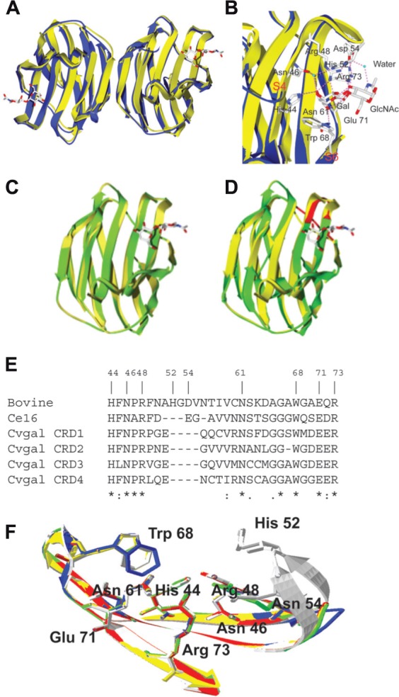
Structures of bovine galectin-1 and Bufo arenarum galectin-1 like protein. (A) The ribbon diagram shows the overlap of the toad (B. arenarum) galectin-1 like protein (blue, PDB 1GAN) and bovine (Bos taurus) galectin-1 (yellow, PDB 1slt) in complex with LacNAc (stick representation). (B) Carbohydrate-binding sites of B. arenarum galectin-1 like protein (blue) and bovine galectin-1 (yellow). The interactions of amino acid residues with LacNAc are shown for the bovine galectin-1. The OH at C4’ of Gal (in LacNAc) makes hydrogen bond interactions with the highly conserved residues His44, Asn46, and Arg48. The OH at C6’ makes similar interactions with the Asn61 and Glu71. Trp68 participates in a stacking interaction with the Gal ring carbons and restricts orientation of the OH at C4’ to the axial form. In GlcNAc moiety of the LacNAc, the hydrogen bond interactions are involved with the protein through the C3-OH with Agr48, Glu71, and Arg73. Additional interactions are involved via a water molecule that bridges the nitrogen of the NAc group with His52, Asp54, and Arg73. (C) Drgal1-L2 (green) was modeled at the SWISS-MODEL Protein Modeling Server (http://swissmodel.expasy.org) based on the bovine galectin-1 structure (yellow, PDB 1slt). All nine residues that form the carbohydrate-binding cassette in mammalian galectin-1 are present in the putative binding site of Drgal1-L2. All side chains of these residues were within 0.5 Å of the equivalent side chains of the bovine galectin-1. (D) C. elegans 16-kDa galectin (Lec-6; shown in green) was modeled at the SWISS-MODEL Protein Modeling Server (http://swissmodel.expasy.org) based on the bovine galectin-1 structure (shown in yellow, PDB 1slt). The model reveals that a shorter loop (indicated by arrow) between strands 4 and 5 is responsible for its unique binding profile. (E) Alignment of bovine galectin-1, Ce16 (C. elegans 16 kDa galectin), and CRD1 to -4 of CvGal. (F) Homology modeling of CvGal CRDs. Bovine galectin-1, CRD-1, -2, -3, and -4 are shown in white, blue, yellow, red, and green, respectively. Numbering of amino acid residues is based on bovine galectin-1.