


Figure 4.
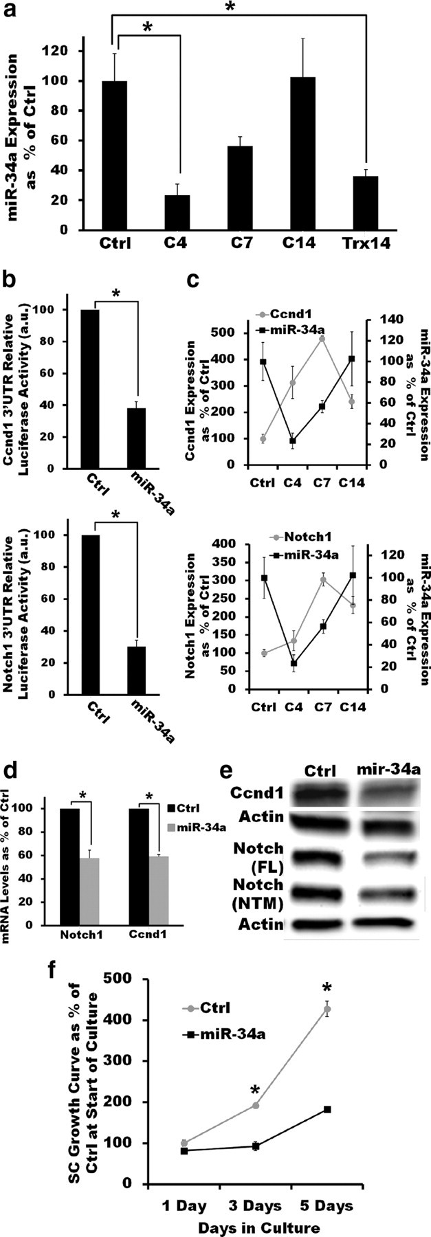
miR-34a targets Notch-1 and Ccnd1 to regulate SC proliferation in vitro. a, The dynamic regulation of miR-34a expression in sciatic nerves after crush injury suggests a potential role in the modulation of cell cycle regulators. miR-34a levels are significantly decreased 4 d after crush injury (p < 0.01, two-tailed Student's t test) but recover as redifferentiation ensues (C14). After nerve transection (Trx), an injury paradigm in which remyelination does not occur, mir-34a levels remain low compared with uninjured nerves even 14 d after injury (p < 0.05, two-tailed Student's t test). Reported values are normalized to U6; error bars represent SEM; n = 3–6 mice at each time point. b, miR-34a directly interacts with the 3′UTRs of Ccnd1 (top) and Notch1 (bottom). Overexpression of miR-34a but not of Ctrl miRNA in HEK293T cells expressing a luciferase reporter construct carrying the 3′UTR of either Notch1 or Ccnd1 results in significantly decreased luciferase activity (p < 0.01 in both cases, two-tailed Student's t test). Error bars represent SEM; n = 3 independent assays c, qRT-PCR measurements show that the expression pattern of Ccnd1 (top) and Notch1 (bottom) after injury is inversely correlated with that of miR-34a, suggesting a direct targeting of these transcripts by miR-34a. Reported values are normalized to GAPDH for mRNA and U6 for miRNAs; error bars represent SEM; n = 3–6 mice at each time point. d, e, Lentivirus-mediated mir-34a overexpression (≈3-fold) in cultured SCs significantly reduces Notch1 and Ccnd1 mRNA (d; p < 0.05, in both cases, two-tailed Student's t test) and protein levels (e, ≈2.5-fold decreas). FL, Full-length; NTM, cleaved transmembrane/intracellular region. qRT-PCR reported values are normalized to GAPDH; error bars represent SEM; n = duplicate wells from three independent assays. f, Lentivirus-mediated mir-34a overexpression (≈3-fold) impairs the ability of SCs to proliferate in the presence of DRG axons. miR-34a-expressing SCs have a significantly decreased axon-induced proliferation in vitro compared with control SCs (no significant difference at day 1; p < 0.01 at days 3 and 5; two-tailed Student's t test). Error bars represent SEM; n = triplicate wells from two independent assays. *p < 0.05 statistical significance.