


Figure 1. Neuroprotective functions of extracellular PGRN.
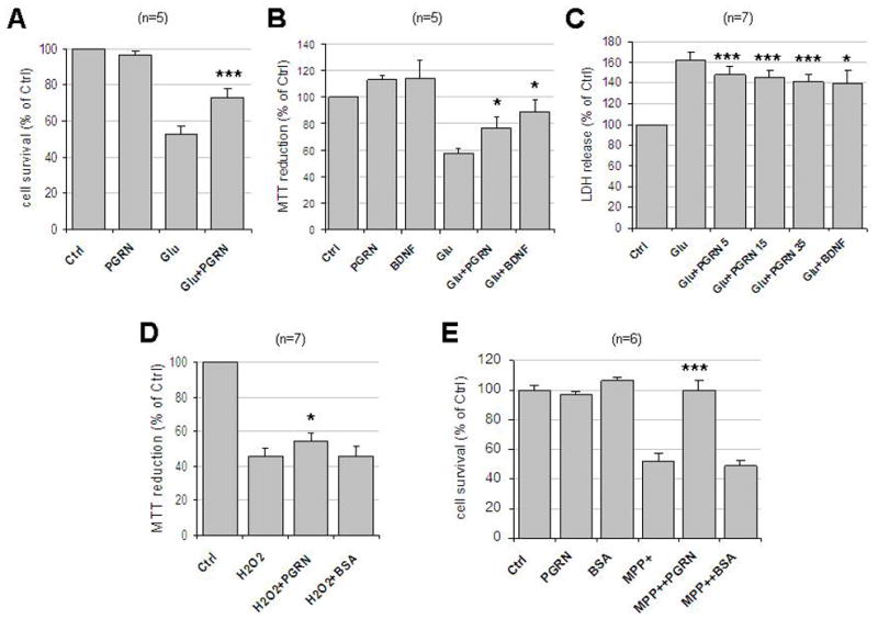
(A) Rat cortical neurons were cultured in 24-well plates in Neurobasal Media plus B27 supplement. At 8–12 DIV, neurons were pre-treated with 35nM PGRN for 30 min followed by 50μM glutamate incubation for 3 hrs. Cells were then fixed with 4% paraformaldehyde and stained with Hoechst 33342. Five pictures were taken from each well and each condition represented the average of four wells. Cell survival was measured by counting the number of cells with normal nuclear morphology. Results (mean ± SE) were calculated from five independent experiments. ***, p<0.005 comparing between cultures treated with glutamate in the presence or absence of PGRN (paired t-test). (B) Cortical neurons as above cultured in 96-well plates were pre-treated with 35nM PGRN or 50ng/ml BDNF for 18 hrs followed by 50μM glutamate for 3 hrs. Cell viability was evaluated by MTT assay and normalized to control (ctrl). Results (mean ± SE) were summarized from five independent experiments and in each experiment each condition is the average of six identical wells. *, p<0.05 comparing between cultures treated with glutamate in the presence or absence of PGRN or BDNF (paired t-test). (C) Neuronal cultures as above were pretreated with different concentrations (5nM, 15nM or 35nM) of PGRN or 50ng/ml BDNF for 2hrs followed by 50μM of glutamate treatment for 24 hrs. The cell-free culture supernatant was collected and LDH release was determined as per Manufacturer’s instructions. Results (mean ± SE) were summarized from seven independent experiments. *, p<0.05; ***, p<0.005 comparing between cultures treated with glutamate in the presence and absence of PGRN or BNDF (paired t-test). Numbers next to PGRN indicate concentrations in nM. (D) Rat cortical neuronal cultures as in (B) were pre-treated with either 35nM PGRN or BSA for 24 hrs followed by 25μM H2O2 treatment overnight. Cell viability was evaluated as in (B) and normalized to control. Results (mean ± SE) were summarized from seven independent experiments and in each experiment each condition is the average of six identical wells. *, p<0.05 comparing glutamate-treated cultures in the presence or absence of PGRN (paired t-test). (E) Neurons in 12-well plates were pre-treated with either 35 nM PGRN or BSA for 24 hrs followed by 40 μM MPP+ treatment for 24 hrs. Cell survival was evaluated by counting the number of intact nuclei in a haemacytometer after lysis (see Materials and Methods). Results (mean ± SE) were summarized from six independent experiments. ***, p<0.005 comparing between cultures treated with glutamate in the presence and absence of PGRN (paired t-test). Ctrl, no treatment; Glu, glutamate.