


Figure 2.
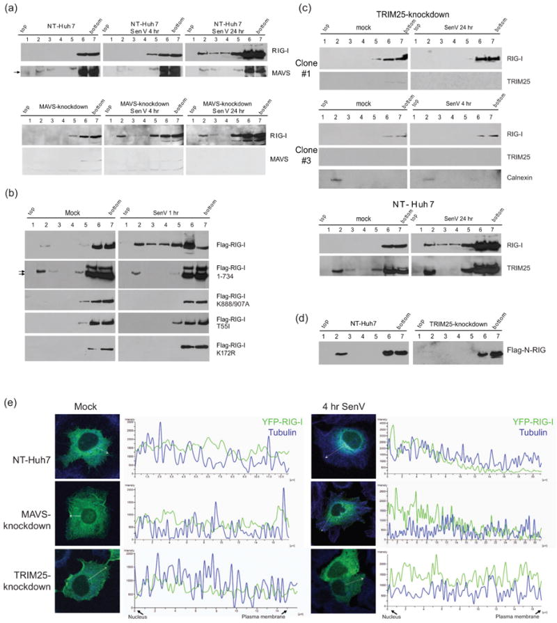
Requirements for RIG-I translocation. (a-d) Protein distribution across membrane flotation gradients was monitored by immunoblot assay. (a) NT-Huh7 and MAVS-knockdown Huh7 cells after 4 or 24 hr SenV infection or mock infection. (b) Distribution of wtor mutant FLAG-RIG-I in Huh7 cells after 1 hr of mock-infection (left) or SenV-infection (right). Arrows denote positions of FLAG-RIG-I 1-734. (c) Distribtion of endogenous RIG-I and TRIM25 from within TRIM25-knockdown (upper) and NT-Huh7 cells (lower). Cells were mock-infected (left) or infected with SenV for 4 or 24 hr prior to harvest. The same blots were stripped and re-probed to detect RIG-I and TRIM25. (d) Distribution of constitutively active FLAG N-RIG mutants expressed in NT control or TRIM25-knockdown Huh7 cells. (e) Distribution of YFP-RIG-I (green) and tubulin (blue) in NT control, MAVS-knockdown, and TRIM25-knockdown Huh7 cells. Mock-infected SenV-infected cells were imaged by confocal microscopy and analyzed for the fluorescence intensity across the sections indicated by white vectors.