


Figure 5.
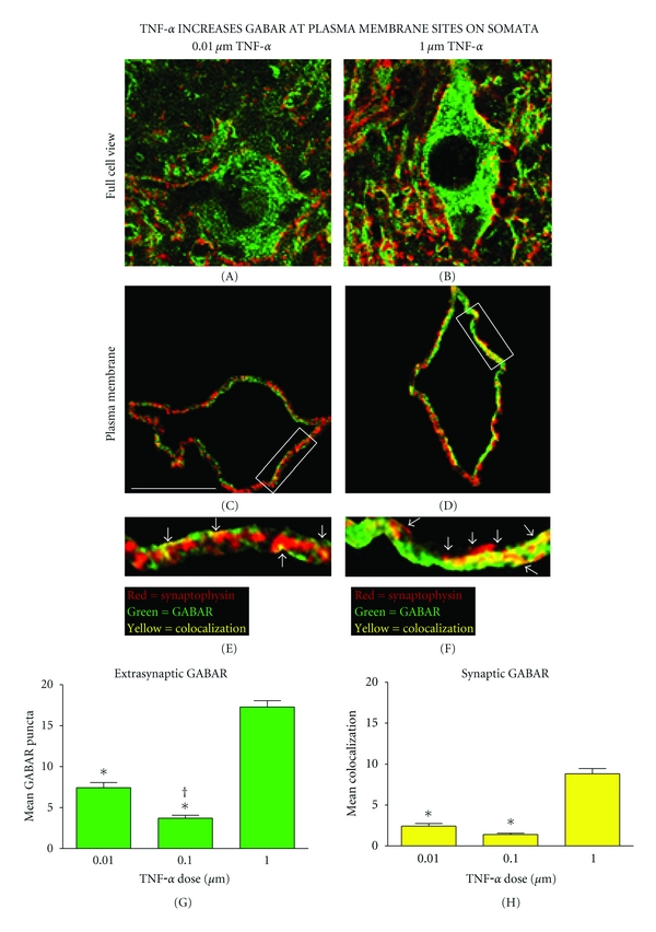
Increased GABAARs on the plasma membrane of spinal neurons after TNFα injection. ((a) and (b)) An image analysis program identified the confocal plane with the greatest colocalization of GABAAR and synaptophysin for each motor neuron. ((c) and (d)) A 2 μm wide cutout of the image containing the somatic plasma membrane. ((e) and (f)) Boxed plasma membrane fractions enlarged to demonstrate representative differences in extrasynaptic (green) and synaptic (yellow) GABAAR puncta on the plasma membrane following the highest and lowest injections of TNFα in vivo. ((g) and (h)) The highest dose of TNFα yielded a significant increase in extrasynaptic and synaptic GABAARs on the plasma membrane (extrasynaptic GABAAR *P < 0.001 from low and middle dose; synaptic GABAAR *P < 0.001 from low and middle dose). Furthermore, the lowest dose of TNFα produced a significant increase in extrasynaptic GABAARs relative to the middle dose († P = 0.002). Bars represent group means across > 800 confocal image stacks (12 subjects, n = 4 subjects per group). Error bars reflect SEM. Scale bar, 30 μm.