


Figure 3.
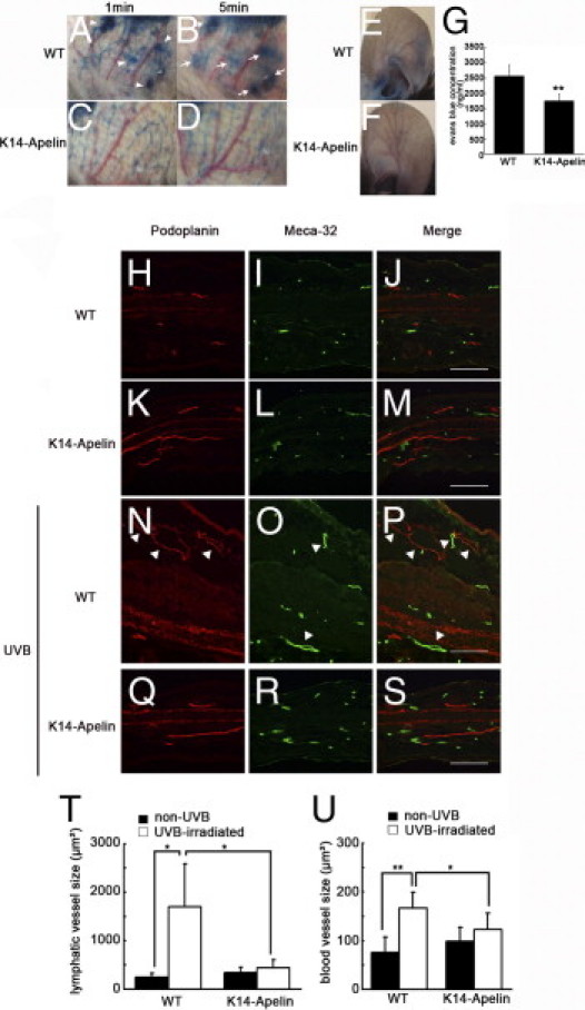
Activation of apelin/APJ signaling inhibited UVB-induced inflammation by blocking enlargement of lymphatic vessels and blood vessels. A–D: Evans Blue dye was injected intradermally into the rim of mouse ears. After 5 minutes, Evans Blue dye had extravasated from lymphatic vessels in UVB-irradiated WT mouse skin (A, arrowheads; B, arrows), but such leakage was attenuated in K14-apelin mice after UVB (C and D). E–G: Miles assay revealed that UVB exposure induced vascular hyperpermeability of WT mice (E), but this effect was markedly inhibited in K14-apelin mice (F). G: Quantitative analysis demonstrated increased Evans Blue leakage in the ear of UVB-irradiated WT mice, compared with UVB-irradiated K14-apelin mice. *P < 0.01. H–S: Double immunofluorescence staining of ear sections for podoplanin (red) and Meca-32 (green) revealed enlargement (arrowheads) of podoplanin-positive lymphatic vessels and Meca-32-positive blood vessels in UVB-irradiated WT mice (N–P), compared with nonirradiated mice (H–M). This enlargement of lymphatic vessels and blood vessels after UVB irradiation was blocked in K14-apelin mice (Q–S). Scale bars: 100 μm. T and U: According to morphometric analyses using IP-LAB software version 4.0, the average size of lymphatic vessels (T) and blood vessels (U) was significantly reduced in K14-apelin mice after UVB irradiation (*P < 0.05). In contrast, size of lymphatic vessels and blood vessels was increased in WT mice irradiated with UVB. *P < 0.05; *P < 0.01.