


Figure 1.
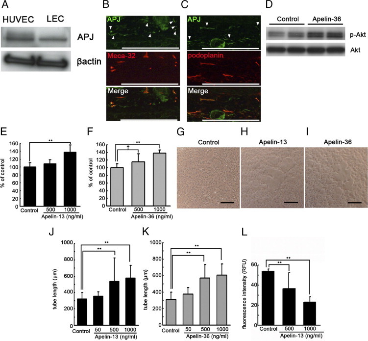
Apelin promotes formation and stabilization of lymphatic endothelial cells (LECs) in vitro. A: Immunoblot analyses confirmed that the apelin receptor APJ was expressed by both LECs and HUVECs. B and C: Double immunofluorescence analyses for APJ (green) and blood vessel marker Meca-32 (red, B) or lymphatic vessel marker podoplanin (red, C) revealed that APJ was expressed by both blood vessels and lymphatic vessels in vivo. Arrowheads show APJ expression in blood vessels (B) and lymphatic vessels (C). D: Treatment of LECs with 1000 ng/mL apelin-36 resulted in increased phosphorylation of Akt, compared with untreated cells. E and F: Apelin-13 (E) and apelin-36 (F) induced migration of LECs in a dose-dependent manner, compared with untreated control cells. G–I: Incubation of LECs with 500 ng/mL or 1000 ng/mL apelin-13 (H and J) and apelin-36 (I and K) enhanced cord formation in a dose-dependent manner after overlay with a type I collagen gel, compared with controls (G). Scale bars: 100 μm. L: The addition of 500 or 1000 ng/mL of apelin-13 decreased the fluorescence intensity of permeated fluorescein isothiocyanate-dextran from LEC, compared with controls. Data are expressed as means ± SD. **P < 0.01; †P < 0.1.