


Figure 6.
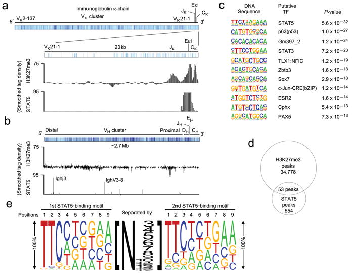
Tetrameric STAT5 binding mediates H3K27 trimethylation in vivo. (a-b) ChIP-seq analysis of Rag2-/- pro-B cell populations expanded in vitro for 2 days in the presence of IL-7 (10 ng/ml). ChIPs were performed using anti-STAT5 and anti-H3K27me3. (a) Schematic of the Igk locus noting the location of Vκ, Jκ, Eκi and Cκ gene segments (mm9 chromosome 6: 70,653,572-70,676,748). The regions of detected H3K27me3 and STAT5 binding are provided. (b) Schematic of the Igh locus noting the location of VH, DH, JH, Eμ, and CH gene segments (mm9 chromosome 12: 114,496,979-117,248,165). The regions of detected H3K27me3 and STAT5 binding are provided. (a,b) Results are presented as a smoothed tag density where tag = sequence read. (c) Conserved de novo DNA sequence motifs identified among STAT5-bound regions. (d) Venn diagram showing the common peaks obtained from STAT5 and H3K27me3 ChIP-seq analysis. (e) Predicted tetrameric STAT5 binding motif determined from the gene regulatory regions demonstrating STAT5 and H3K27me3 common peaks (Supplementary Tables 3,4). The size of the letter at each position represents the percentage of presence of that nucleotide in that position. The size of the number represents the number of the nucleotides present between two STAT5 binding motifs.