


Figure 2.
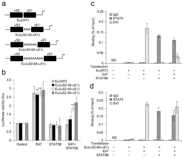
STAT5 and E2A compete for binding at their respective Eκi sites. (a) Schematic representation of the 3, 6 and 9 adenine oligonucleotide spacers (between κS2 and κE1) expressed in pGL3-promoter luciferase plasmids. (b) Cos7 cells were transfected with the pGL3-promoter luciferase reporter plasmids described in (a) along with plasmids encoding pRL-TK (Renilla luciferase) and indicated molecules or appropriate control plasmids. After 48 h, dual luciferase assays were performed on cell lysates (average ± s.d, n=3). *P<0.001, versus Eκi(WT) control; ** P<0.001, versus Eκi(κS2-3A-κE1) control; *** P<0.001, versus Eκi(κS2-6A-κE1) control; †P<0.001, versus Eκi(κS2-9A-κE1) control. (c,d) ChIP assay of nuclear preparations from Cos7 cells transfected as in (b) with (c) Eκi(WT) or (d) Eκi(κS2-9A-κE1)(average ± s.d, n=3). ChIPs were performed with antibodies specific for STAT5, E47 or control IgG1 and assayed with PCR primers specific for the Eκi fragment in the reporter plasmids. *P<0.001, versus E47 binding in only E47 expressed cells.