


Fig. 3.
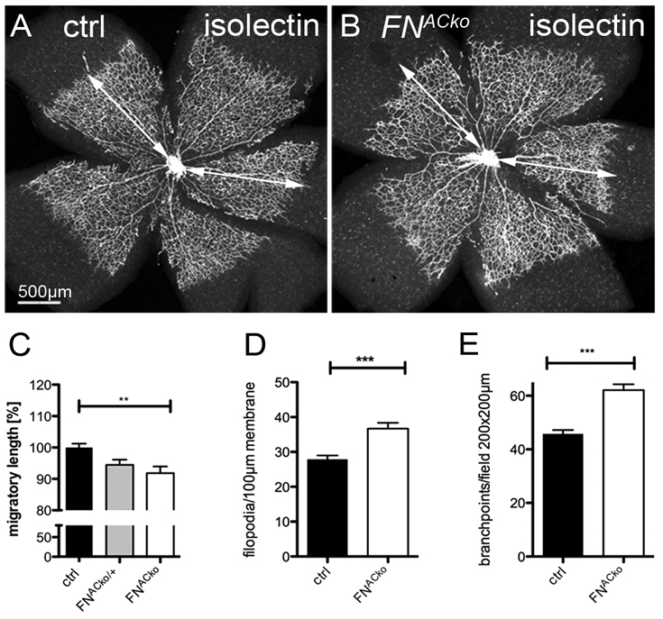
Loss of astrocytic FN results in reduced migration of vessels. (A,B) Visualization of the retinal vasculature (P5) by isolectin-B4 immunofluorescence staining revealed reduced migration towards the retinal periphery in mice deficient of astrocytic FN. Vessel migration was quantified by measuring total length as illustrated by the white arrow (A,B). (C) Radial expansion of superficial vessels at P5 in control (n>5), heterozygous (FNACko/+, n>3) and FNACko mice (n>5). **P<0.01 one-way ANOVA. Values represent mean+s.e.m. (D) The number of filopodial protrusions per vessel length at the migrating vascular front of P5 mice (n≥20). ***P<0.0001. Values represent mean+s.e.m. (E) The number of branch points per image field of P5 FNACko and control mice, n≥10. ***P<0.0001. Values represent mean+s.e.m.