


Figure 1.
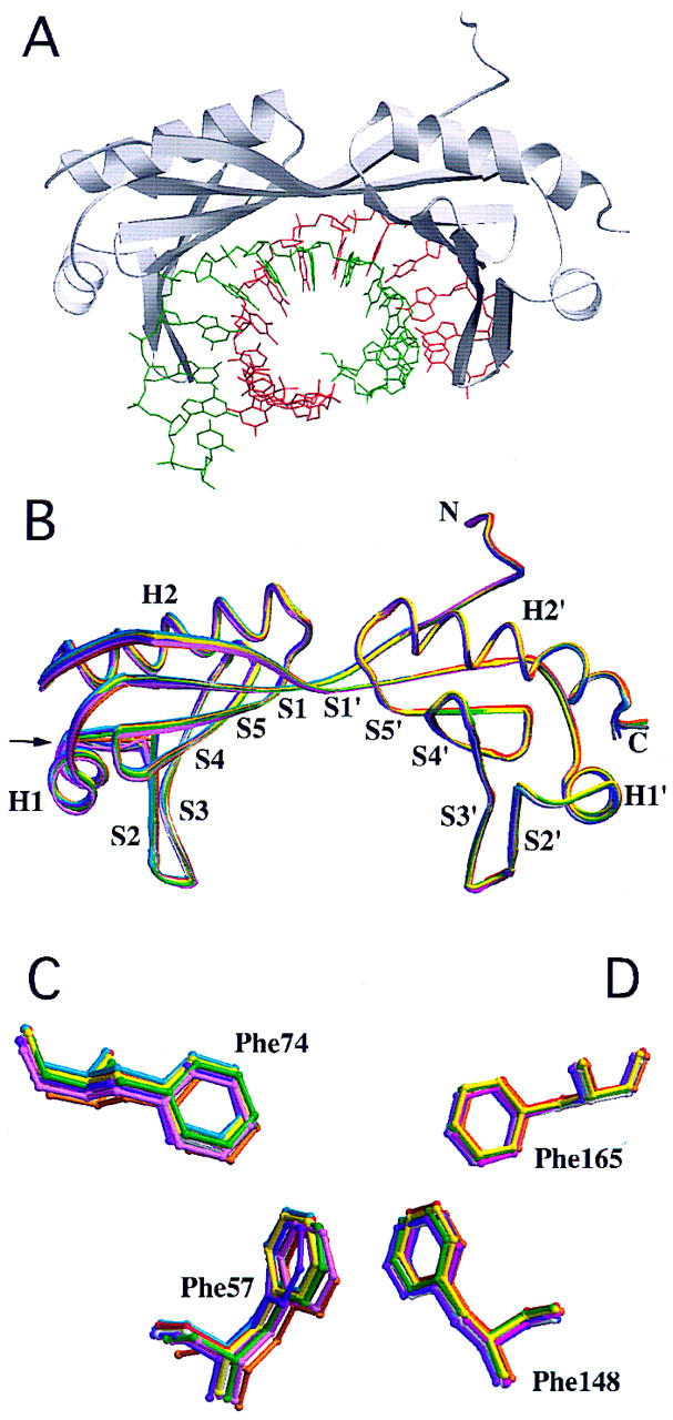
Structure of TBP bound to the TATA element. (A) MOLSCRIPT drawing of the TBP–TATA box complex (coding strand shown in green) viewed perpendicular to the twofold quasisymmetry axis. (B) Overlay of the polypeptide backbones of 11 TBP–DNA complexes, by use of a spaghetti representation with a different color for each cocrystal structure and the view shown in A. Atomic stick figure representations of the amino-terminal (Phe-57 and Phe-74) (C) and carboxy-terminal (Phe-148 and Phe-165) (D) phenylalanine pairs from the 11 TBP–DNA complexes, by use of the color coding in B. Overlay was performed by least-squares superposition of the α-carbons only.