


Figure 2.
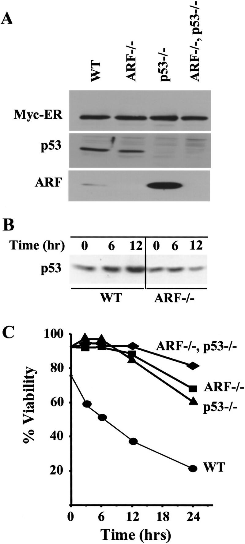
ARF and p53 mediate c-Myc-induced apoptosis in primary B cells. (A) Levels of Myc–ERTM, p53, and ARF in wild-type, ARF−/−, p53−/−, and ARF–p53 doubly null pre-B cells infected with a retrovirus encoding Myc–ERTM–GFP. Infected pre-B cells sorted for green fluorescence were expanded, lysed, and equal quantities of protein were assessed by immunoblotting with antibodies specific for each protein. (B) Kinetics of p53 induction in response to 4-HT in the same experiment shown in C. p53 was detected by immunoblotting as in A. No signals were detected in lysates of cells lacking p53 (negative data not shown). (C) Steady-state levels of apoptosis in the indicated primary pre-B cells at 14 days after infection are indicated at the 0 hr time point. 4-HT was added to the indicated primary pre-B cell cultures to activate Myc–ERTM, and their viability was determined at intervals thereafter by trypan blue dye exclusion and confirmed by analysis of subdiploid DNA content or staining with Hoescht 33342. The data are representative of three independent experiments.