


Figure 3.
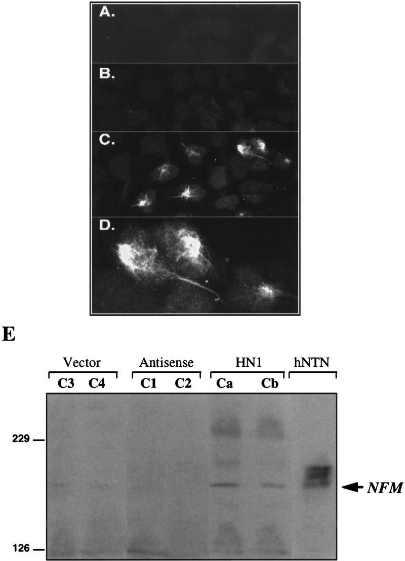
Expression of NF-M protein in cells stably transfected with Hel-N1. Confocal microscopy images of vector-transfected cells (A), antisense-Hel-N1-transfected cells (B), and Hel-N1-transfected cells immunostained with anti-NF-M antibody(C,D). (D) Magnified top right corner of C. (E) Immunoblot stained with anti-NF-M antibody. Total cell extracts (150 μg) from two vector-transfected clones (C3 and C4), two antisense-Hel-N1-transfected clones (C1 and C2), two Hel-N1-transfected clones (Ca and Cb), and untransfected differentiated neurons (hNTN) were loaded onto 6% acrylamide gels and analyzed for NF-M expression by ECL immunoblotting.