



Abstract
Platelets are a remarkable mammalian adaptation that are required for human survival by virtue of their ability to prevent and arrest bleeding. Ironically, however, in the past century, the platelets’ hemostatic activity became maladaptive for the increasingly large percentage of individuals who develop age-dependent progressive atherosclerosis. As a result, platelets also make a major contribution to ischemic thrombotic vascular disease, the leading cause of death worldwide. In this brief review, I provide historical descriptions of a highly selected group of topics to provide a framework for understanding our current knowledge and the trends that are likely to continue into the future of platelet research. For convenience, I separate the eras of platelet research into the “Descriptive Period” extending from ~1880-1960 and the “Mechanistic Period” encompassing the past ~50 years since 1960. We currently are reaching yet another inflection point, as there is a major shift from a focus on traditional biochemistry and cell and molecular biology to an era of single molecule biophysics, single cell biology, single cell molecular biology, structural biology, computational simulations, and the high-throughput, data-dense techniques collectively named with the “omics” postfix. Given the progress made in understanding, diagnosing, and treating many rare and common platelet disorders during the past 50 years, I think it appropriate to consider it a Golden Age of Platelet Research and to recognize all of the investigators who have made important contributions to this remarkable achievement.
Introduction
Platelets are a remarkable mammalian adaptation that are required for human survival by virtue of their ability to prevent and arrest bleeding. Ironically, however, in the past century, the platelets’ haemostatic activity became maladaptive for the increasingly large percentage of individuals who develop age-dependent progressive atherosclerosis. As a result, platelets also make a major contribution to ischaemic thrombotic vascular disease, the leading cause of death worldwide [1]. Thus was born the need to develop anti-platelet therapies to attenuate platelet function in individuals at risk of arterial thrombosis. The wide range of platelet contributions to health and disease has stimulated intense study of platelet function. In this brief review, I provide historical descriptions of a highly selected group of topics to provide a framework for understanding our current knowledge and the trends that are likely to continue into the future of platelet research. For convenience, I will separate the eras of platelet research into an initial “Descriptive Period” extending from ~1880–1960, during which many of the classic clinical features of platelet disorders were detailed (Figure 1) and a subsequent “Mechanistic Period” encompassing the past ~50 years, made possible by the introduction of biochemical, cell biologic, molecular biologic, and most recently, structural biologic, genomic, and computational techniques. Unfortunately, the limitations of space make it impossible to include the names in the text of the large number of distinguished investigators who made many of the important contributions. Several excellent histories of platelet discoveries are referenced throughout for those interested in additional details.
Figure 1.

Timeline of First Clinical Reports of Important Platelet Disorders
*When multiple dates are provided, they reflect different important descriptions of the disorder.
The Descriptive Period
Discovery of Platelets and Megakaryocytes
The Descriptive Period began with the elegant and comprehensive intravascular microscopy and ingenious in vitro flow chamber studies reported by Bizzozero in 1881–2 [2, 3]. Although others probably observed platelets earlier [4, 5], he correctly identified the platelet’s role in both haemostasis and thrombosis. He was also the first to describe bone marrow megakaryocytes [6], but Wright was the first to identify the megakaryocyte as the precursor cell to the platelet, aided by the new staining techniques he developed [7, 8]. Osler in 1886 established that platelets contribute to human thrombotic disorders, discovering them in white thrombi in atheromatous aortic lesions and on diseased heart valves [9].
Descriptions of Clinical Syndromes and Early Studies of Platelet Physiology
Many important clinical disorders were described during the Descriptive Period, providing compelling evidence of the importance of platelets in haemostasis and spurring interest in platelet physiology [10–13]. These include what would later be renamed immune thrombocytopenia (1735/1883) [14], May-Hegglin anomaly (1909/1945) [15], thrombocytopenic haemorrhage (1910) [16], Glanzmann thrombasthenia (1918) [17], thrombotic thrombocytopenic purpura (Moschcowitz syndrome, 1924) [18, 19], von Willebrand disease (1926) [20–22], and Bernard-Soulier syndrome (1948) [23]. Similarly, assays for platelet function were developed, including clot retraction (1878/1951) [24] and the Duke bleeding time (1910) [16]. Histologic examination of sites of vascular injury in experimental animals using light microscopy and later electron microscopy established the sequence of events, including platelet adhesion and aggregation, followed by degranulation, the loss of distinct borders between platelets, and platelet thrombus contraction, which were collectively termed “viscous metamorphosis” [5, 25]. Studies of serum led to the discoveries that thrombin is a strong platelet activator and that platelets are secretory cells that store and release the vasoactive compound serotonin, a derivative of tryptophan [26]. The observation that platelets adhere to connective tissue led to the discovery that collagen is also a potent platelet activator [25, 27]. Clot retraction was studied in detail [24] leading to the discovery that platelets contain the contractile proteins actin and myosin, initially termed thrombosthenin [28], the first non-muscle cell shown to have these elements [29].
The different mechanisms of thrombocytopenia were studied intensely during the Descriptive Period. In particular, the pioneering and courageous studies of Harrington and colleagues, in which blood components from patients with immune thrombocytopenia were infused into volunteers, including Harrington himself, demonstrated that the agent causing immune thrombocytopenia could be passively transferred with plasma [30]. His group went on to define many important aspects of immune thrombocytopenia, including the crucial role of antibodies, and the beneficial effects of splenectomy and glucocorticoid therapy on platelet clearance [31].
Erich von Willebrand described the bleeding disorder that bears his name based on studying a 5 year old girl in the Åland Islands and 65 other members of her family [20–22]. The disorder clearly differed from haemophilia in its cardinal manifestations since it was inherited as an autosomal dominant trait rather than as an X-linked trait, bleeding was primarily mucocutaneous, the bleeding time was prolonged, and the clotting time was normal. Thus, the disorder had many of the characteristics of a platelet abnormality, but it was unclear whether the defect was intrinsic to the platelet or resided in a plasma cofactor needed for platelet function. Studies conducted in the 1950s by several groups demonstrated a partial deficiency in factor VIII, but when compared to the levels in patients with haemophilia A, the reduction was not able to account for the bleeding symptoms [32–36]. They also demonstrated that the plasma fraction termed Cohn fraction 1 from normal individuals could correct the bleeding time when infused into patients with von Willebrand disease and produce a paradoxically exaggerated increase in factor VIII [32, 33].
Early Treatments
A number of important therapies were developed during the Descriptive Period. Platelet transfusion therapy for thrombocytopenia was inaugurated with Duke’s compelling demonstration in 1910 of the immediate haemostatic efficacy of direct donation of whole blood to correct severe thrombocytopenia [16]. It would take another 35 years, however, before there was a systematic effort to improve the techniques of platelet transfusion, spurred by the recognition that thrombocytopenic haemorrhage was a major cause of death after radiation exposure to atomic weapons [37, 38]. Further impetus for improving platelet transfusion therapy near the end of the Descriptive Period came from the evidence that thrombocytopenic haemorrhage was also a major cause of death from radiation therapy and the then-new chemotherapeutic agents [39]. Thus, platelet transfusion therapy became crucial for the success of modern chemotherapy. Splenectomy for what would later be termed immune thrombocytopenia was introduced in 1916 based on the recommendation of Kaznelson, who was a medical student in Prague [14, 40] (Figure 2). The first surgery was performed by Professor Schloffer and the first patient’s platelet count increased from less than 1,000/μl to ~500,000/μl. Corticosteroid therapy was also introduced for immune thrombocytopenia during the Descriptive Period [41].
Figure 2.
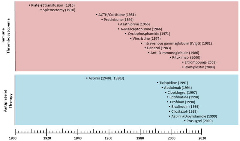
Dates of Introduction or U.S. FDA Approval of Selected Therapies for Platelet Disorders
The Mechanistic Period
Megakaryopoiesis and Thrombopoiesis
Although the existence of a humoral factor that stimulates megakaryocyte production was postulated by Kelemen near the end of the Descriptive Period based on rodent studies of thrombocytopenia [42], attempts to purify thrombopoietin (Tpo) during the next 30 years achieved only limited progress [43, 44], in part because they relied on complex in vivo, radiolabeled methionine incorporation assays [45]. In vitro culture methods on semisolid media developed in the 1970s made it possible to define the colony- and burst-forming megakaryocyte units (CFU-MK and BFU-MK), along with their precursors, as well as variably-specific transcription factors (e.g., SCL, GATA1, GATA2, NF-E2, and ets family members) and cytokine receptors involved in megakaryopoiesis [45]. The breakthrough, however, came when several groups recognised that the cellular proto-oncogene of the murine myeloproliferative leukaemia virus (c-Mpl) was a member of the cytokine receptor family, and that because it was expressed in megakaryocyte precursors, it was a strong candidate to be the receptor for Tpo. In 1994, studies by multiple groups using c-MPL for affinity purification or direct biochemical purification techniques resulted in the purification and cloning of Tpo [46–49]. Tpo was shown to increase platelet counts in animals and humans by enhancing both megakaryocyte proliferation and survival. Loss of either Tpo or its receptor was shown to result in severe thrombocytopenia, demonstrating Tpo’s important physiologic role. In fact, a number of patients with inherited amegakaryocytic thrombocytopenia were found to have mutations in c-MPL [50].
Studies of Tpo also provided insights into the regulation of the platelet count, which is normally maintained at a level that is ten-fold higher than that required to prevent spontaneous bleeding. Much can be explained by a model in which Tpo catabolism is controlled by the total mass of platelets and megakaryocytes via their binding and degradation of Tpo [51], with potential additional roles for regulated Tpo expression and/or cytokine (e.g., IL-6)-induced upregulation of Tpo expression [45].
Studies of megakaryopoiesis have also provided insights into the endomitotic process that allows megakaryocytes to increase their ploidy and mass without undergoing division [52–55]. They also shed light on how organelles and granules move along microtubule tracks to the tips of proplatelets [56, 57] before they extend into the vascular sinuses in the bone marrow, bend and fragment, giving rise to thousands of platelets [58–61]. Studies of the myeloproliferative and megakaryocytic disorders associated with Down’s syndrome trisomy 21 have provided valuable insights into transcription factors that control megakaryopoiesis [62, 63].
Platelet Biochemistry
The early phase of the Mechanistic Period was marked by bringing the new tools of biochemistry to bear on the platelet phenomena that had already been described and by developing more quantitative techniques to evaluate and dissect the phenomena. The platelet’s protein and lipid composition, inorganic constituents, enzymatic activities, and energy metabolism were defined and quantified [10, 64]. Hellem and his colleagues developed a method for measuring platelet “adhesiveness” using columns of glass beads, and found an association between “adhesiveness” and the blood hematocrit [65]. Based on that finding, they postulated that red cells must have a powerful platelet activating agent and this led them to discover the platelet aggregating activity of ADP [66]. Modifications of the platelet “adhesiveness” assay (later termed the platelet retention test since it reflects both adhesion to glass and platelet aggregation) helped to define the abnormalities in platelet function in uraemia, afibrinogenaemia, Glanzmann thrombasthenia, Bernard-Soulier syndrome, and von Willebrand disease, including the accentuation of the platelet function defect in von Willebrand disease under high shear conditions [67–69]. It also furnished the first assay to support the purification of von Willebrand factor (VWF) [70]. Biochemical analysis of platelet granule contents demonstrated the presence of adhesive glycoproteins such as fibrinogen, vWf, and thrombospondin-1 in α granules, along with cytokines and platelet-specific proteins such as platelet factor 4 and the β-thromboglobulin family of proteins [71–73]. The presence of a “storage pool” of ADP, ATP, and serotonin in the calcium-rich dense bodies was also discovered and characterised [74].
The Aggregometer and Flow Chamber Studies
The need for more quantitative and robust methods of assessing platelet function led both Born and O’Brien to develop turbidometric platelet aggregometry in 1962 [75, 76]. This allowed for the characterisation of the unique platelet effects of the different agonists that had been, and were being, discovered, including ADP, epinephrine, thrombin, collagen, vasopressin, and serotonin. The discovery that with citrated platelet-rich plasma ADP and epinephrine produce aggregometer tracings that could be divided into two separate phases, or waves, led to the definition of the role of the platelet release reaction, including the release of ADP from dense bodies and the release of arachidonic acid and its subsequent conversion into thromboxane A2, in amplifying the platelet aggregation response [77–80]. The subsequent development of the lumi-aggregometer allowed for simultaneous measurement of both platelet aggregate formation and release of ATP from dense bodies [81]. Aggregometer studies were also crucial in more precisely defining the roles of phospholipids, phosphoinositol hydrolysis, diacylglycerol, and Ca2+ mobilisation in platelet activation [82–86], as well as the functional defects in many of the previously described inherited platelet disorders [87–90], and the newly described Storage Pool disorders [91], the Gray platelet syndrome [92], and disorders of platelet secretion [93, 94]. The roles of fibrinogen and VWF in mediating platelet-platelet interactions via binding to αIIbβ3 [95–98] and GPIb [99], respectively were also facilitated by aggregometer studies. Similarly, studies using newly designed flow chambers defined platelet-ligand and platelet-platelet interactions as a function of shear rate, introducing and partially simulating an important aspect of the in vivo milieu [100–103].
Platelet Coagulant Activity
Platelets were shown to greatly accelerate blood coagulation and this property was initially termed platelet factor 3 (PF-3) [10, 104]. This factor was only “made available” when platelets were activated under certain conditions, or when they were subjected to physical trauma, as with freezing and thawing. Extensive studies of the platelet lipids and proteins responsible for the nearly 100,000-fold increase in the rate of thrombin generation identified exteriorisation of the negatively charged phospholipid phosphatidylserine as a primary contributor to recruitment of coagulation factors and enhancement of catalytic efficiency [105, 106]. A series of platelet protein receptors for coagulation factors were also identified and their contributions continue to be the subject of study [107–109]. The role of fibrin in enhancing platelet-mediated thrombin generation was also identified [110]. Platelet microparticles, small fragments of platelets that circulate in health and in disease, were found to have coagulant activity and to be biologically active [111, 112]. Studies of a single patient, Mrs. Scott, first described by Weiss and colleagues in 1979 [113] provided important insights. Her bleeding symptoms differed from patients with qualitative or quantitative defects in platelets in that she did not have easy bruising; she did, however, have menorrhagia and post-partum haemorrhage. Her platelets failed to expose negatively-charged phospholipids upon activation, apparently as part of generalised defect in cell vesiculation [114–116]. Information from advanced biophysical tools and structural biology insights have been combined to provide a detailed image of the underlying mechanisms by which platelets contribute to thrombin generation [111, 112, 117–120].
The Endothelial Lining, Inhibitors of Platelet Function, and Platelet-Leukocyte Interactions
The endothelial lining of blood vessels prevents platelet interactions with the vessel wall by both synthesising platelet inhibitors and “hiding” the subendothelial platelet-adhesive proteins from the platelet’s view. Studies by the groups led by Moncada and Weksler demonstrated that endothelial cells synthesise prostaglandin I2 (PGI2, prostacyclin) from arachidonic acid [121, 122].
Subsequently, endothelial cells were found to synthesise the platelet inhibitor nitric oxide (NO), which acts synergistically with prostacyclin [123–125]. Finally, Marcus and colleagues also identified an endothelial ecto-ADPase [CD39; ecto-nucleotide pyrophosphatase/phosphodiesterase (E-NPP)] capable of rapidly metabolising the platelet activator ADP (as well as ATP) to the inactive compound AMP [126, 127]. The subsequent conversion of AMP to adenosine by the ecto-5’-nucleotidase CD73 on the endothelial cells of some vessels and on some leukocytes yields adenosine, an inhibitor of platelet activation [128–130]. Marcus also identified the extraordinary high content of arachidonic acid in platelets and the complex transcellular eicosanoid metabolism that occurs between platelets, endothelial cells, and leukocytes [131, 132].
Platelets can also interact with, and roll on, intact endothelium via multiple receptors when the endothelium is activated to secrete large VWF multimers and expose P-selectin [133]. Complex mechanisms involving similar interactions underly platelet-leukocyte interactions [134–136]. When the endothelial cell lining is disrupted, platelets adhere to the denuded surface via receptors for the adhesive proteins that are exposed and/or deposited from plasma, in particular, the interactions of platelet GPIbα with VWF [137–139] and both GPVI and α2β1 with collagen [140, 141]. GPIbα has a carbohydrate-rich region that allows it to extend far from the platelet surface and so it attaches first. Engagement of these receptors initiates platelet activation, resulting in activation of αIIbβ3 and recruitment of additional platelets.
Platelet Receptors and Ligands
The concept of cell receptors controlling cell physiology also emerged in the early part of the Mechanistic Period. Initially receptors were characterised functionally and pharmacologically based on biochemical responses and later they were characterised by their ability to release intracellular Ca++, monitored by cation-sensitive dyes [142, 143]. The introduction of radionucleotides in biomedical research made it possible to perform direct ligand binding studies in select cases [95, 96]. The introduction of sodium-dodecyl sulfate polyacrylamide gel electrophoresis (SDS-PAGE) allowed for detailed biochemical assessment and was a great aid to receptor purification and characterisation [99, 144]. It also provided the initial glycoprotein (GP) nomenclature for many of the receptors [145]. The sensitivity and specificity of SDS-PAGE was greatly enhanced by combining it with platelet surface labelling techniques and immunoblotting [145, 146]. Other immunologic techniques, including cross-immunoelectrophoresis allowed for assessing protein-protein interactions under non-denaturing conditions, helping to define ligand-receptor interactions [99, 144, 145, 147]. Monoclonal antibody development was an additional major advance, providing essentially unlimited amounts of standardised reagents that could unequivocally identify specific receptors by immunoblotting and other techniques [147, 148]. In addition, the development of monoclonal antibodies that could inhibit platelet function provided tools that could dissect the contribution of each receptor [147, 149–152]. This was particularly important in understanding the roles of receptors that could bind more than one ligand, such as αIIbβ3 and αVβ3, and of ligands like VWF and fibrinogen that could bind to more than one receptor. Monoclonal antibodies that reacted selectively with activated αIIbβ3, most notably PAC1 [153], or with P-selectin [154, 155], which is confined to α-granule membranes, made it possible to monitor the activation state of αIIbβ3 and the release reaction, respectively. Similarly, monoclonal antibodies were developed that reported on the conformational changes in receptors after ligand binding [156]. Many of the initial monoclonal antibody studies were performed with radiolabeled antibodies, which required special care and handling. The development of flow cytometry made it possible to substitute more generally available fluorescently-labelled antibodies and permitted analysis of the binding of antibodies to individual platelets [157–159] and even to small platelet microparticles, which are biologically active and support blood coagulation [160, 161].
The introduction of molecular biologic techniques allowed for the detailed analysis of the subunit structures and homology of different receptors, defining the major families of receptors as integrins (αIIbβ3, αVβ3, α2β1, α5β1, and α6β1), leucine-rich repeat receptors (GPIbα, GPIbβ, GPV, GPIX), immunoglobulin repeat receptors (PECAM-1, GPVI, FcγRIIA, CLEC-2), selectins (P-selectin), tetraspanins (CD9, CD151, TSSC6, CD63, and Tspan9) [162, 163], and G protein-coupled 7 transmembrane receptors (including, among others, PAR-1, PAR-4, P2Y1, P2Y12, vasopressin V1a, serotonin 5-HT2A, and adenosine receptors A2a and A2b) [128, 164, 165]. Of particular importance in integrin receptor biology was the extrapolation from studies of α5β1 [166] that both αIIbβ3 and αVβ3 could be inhibited by peptides containing the Arg-Gly-Asp (RGD) sequence [167]. Structural biology studies using nuclear magnetic resonance, x-ray crystallography, electron microscopy, and molecular dynamics simulations now permit atomic level resolution of the receptors and receptor-ligand interactions [168–173]. Transgenic mice with targeted deletions of receptors or mutant receptors have confirmed and extended our understanding of the roles of the different receptors [174, 175], although extrapolation from mouse to human platelets needs to be tempered because of the unique aspects of human versus mouse platelets, such as the repertoire of functional thrombin receptors [176], and the differences in physiology, most notably the mouse’s ~600 beats per minute heart rate!
Insights into the signal transduction pathways triggered by ligand binding also derived from studies of the receptors. Our understanding of collagen signalling through GPVI had its origin in the 1989 description of a patient with an isolated defect in collagen-induced platelet aggregation whose platelets also had a defect in GPVI [177, 178]. Subsequent functional studies and the cloning of the GPVI led to the recognition that it is coexpressed with the FcR γ-chain and requires a dimeric structure to achieve high affinity for collagen [179]. GPVI cross-linked by polymerised collagen (or collagen-related peptides or the snake venom convulxin) was elegantly shown to initiate Srk kinase-dependent tyrosine phosphorylation of the FcR γ-chain ITAM, leading to recruitment and activation of Syk. Downstream from Syk is a cascade of intermediates similar to those in immune cell signalling, including LAT, SLP-76, PI3-kinase and PLCγ2, ultimately leading to the conversion of PIP2 into 1,2-diacylglycerol and inositol 1,4,5-trisphosphate IP3, resulting in an increase in cytoplasmic calcium and granule exocytosis [180]. Signalling through the GPIb/X/V complex leading to inside-out signalling could be inferred from the contribution of αIIbβ3 to ristocetin-induced platelet aggregation and from studies identifying the roles of VWF and fibrinogen in shear-induced platelet aggregation [181]. The signal transduction details, including the roles of tyrosine kinases, were then worked out in several laboratories [182–185]. Signalling through αIIbβ3 (“outside-in”) was shown to include roles for Src, Syk, Gα13 and the phosphorylation of two tyrosine residues in the β3 integrin subunit cytoplasmic tail [186–190]. “Inside-out” signalling to activate αIIbβ3 via changes in its conformation so that it adopts a high affinity ligand binding state was elucidated by elegant functional studies of the roles of talin and kindlin in binding to the β3 cytoplasmic tail and the separation of the transmembrane domains [191–199]. The physiologic role of kindlin-3 was supported by the identification in 2009 of patients with a profound defect in platelet function as part of leukocyte adhesion deficiency-III (LAD-III) resulting in a serious bleeding diathesis in association with mutations in kindlin-3 [197, 198, 200–202]. CLEC-2, a C-type lectin receptor, has multiple ligands including the snake venom rhodocytin, tumor-associated podoplanin and HIV-1, and has been implicated in haemostasis, thrombosis, tumor metastases, and lymphangiogenesis; it also signals through tyrosine phosphorylation of its cytoplasmic tail followed by the binding of Syk [203–208]. Signalling through the G protein-coupled 7 transmembrane domain receptors was investigated in great detail including the pairings with specific Gα subunits, the downstream effects on second messengers, and the feedback control mechanisms [164, 165]. Balancing the platelet receptors involved in initiating signalling are inhibitory receptors containing the immune receptor tyrosine-based inhibitory motif (ITIM), most notably PECAM-1 and G6b-B [209–214]. A third platelet ITIM containing receptor, TREM-like Transcript-1, however, may not serve an inhibitory function; in fact, it may enhance platelet function and contribution to platelet-leukocyte interactions [215].
Clinical Diagnosis, Carrier Detection, and Prenatal Diagnosis
Biochemical and molecular biological advances, most notably the application of the reverse polymerase chain reaction (PCR) to study the small amount of platelet mRNA [216], revolutionised the diagnosis and characterisation of patients with disorders affecting platelet receptors. In particular, Glanzmann thrombasthenia was defined as a defect in either αIIb or β3 [217, 218], Bernard-Soulier syndrome was defined as due to an abnormality in GPIbα, GPIbβ, or GPIX [219], platelet-type von Willebrand disease was defined as a defect in GPIb [220], and other defects were identified in P2Y12, GPVI, and many other receptors [221]. Monoclonal antibodies were rapidly applied to the diagnosis of the disorders and in select cases, carrier detection; they also made possible prenatal diagnosis based on fetal blood sampling at ~18–20 weeks of gestation [222, 223]. Since megakaryocytes also synthesise VWF, platelet mRNA-based PCR was also used to define the molecular biologic defects in VWF and correlate them with the current subtyping of the disorder [224, 225]. Practical benefits to patients accrued rapidly from the advances in molecular biology, including the ability to perform unequivocal carrier detection and prenatal diagnosis using chorionic villus sampling at just ~11 weeks of gestation [226].
The molecular biologic tools also revolutionised our understanding of the alloimmune thrombocytopenias, including neonatal alloimmune thrombocytopenia and post-transfusion purpura, which had been characterised serologically in the 1960s [227–229]. Thus, the amino acid substitutions in the glycoproteins responsible for antibody production were defined, tools were developed to test for potential incompatibilities, and effective therapies introduced [230–235].
von Willebrand Disease
Building on the observation that the clinical platelet function abnormality in von Willebrand disease could be dramatically improved by a plasma factor [32, 33, 236], Ted Zimmerman in Oscar Ratnoff’s laboratory established that most patients with von Willebrand disease have reduced levels of a protein present in normal plasma and the plasma of patients with haemophilia A [237]. A series of biochemical studies ultimately showed that VWF and factor VIII are separate molecules, but as was suspected from the transfusion studies, they circulate as a complex in plasma [22, 238]. The unfortunate choice of the term “factor VIII-related antigen” for VWF created considerable confusion, some of which remains for those new to the field to this day. Studies of VWF immunology, biochemistry, and molecular biology defined its subunit structure, its remarkable multimeric structure, its association with factor VIII, its ability to bind to collagen, GPIbα, and αIIbβ3, and its cleavage by ADAMTS13 [139, 239–242]. Studies of VWF carbohydrate defined functional variants [243] and the role of blood group carbohydrates in its in vivo survival, leading to close correlations between blood type and VWF plasma levels that also translate into haemorrhagic and thrombotic risk [244–247]. The important roles of endothelial cells and megakaryocytes in VWF synthesis were also defined, and the importance of platelet versus plasma VWF debated [248, 249]. The remarkable discovery in 1971 by Howard and Firkin, who systematically studied drugs that produced thrombocytopenia in humans, that the antibiotic ristocetin could agglutinate/aggregate platelets only in the presence of VWF provided more robust functional assays than the platelet retention test for diagnosing von Willebrand disease, purifying VWF, and demonstrating that the higher molecular weight multimers are functionally more effective in agglutinating/aggregating platelets [22, 250–252]. Subsequent studies showed that the snake venom protein botrocetin could also agglutinate platelets in the presence of VWF, providing another tool for assaying VWF [253]. The important role of ADAMTS13 in controlling the size of VWF multimers through proteolysis was later defined, spurred by research on thrombotic thrombocytopenic purpura [19]. Collectively, these advances allowed for the classification of von Willebrand disease into a large number of different subtypes, including the rare platelet-type associated with abnormalities in GPIbα, with both diagnostic and therapeutic importance [240]. Studies of fibrinogen defined the importance of the γ chain C-terminal peptides and other regions in interacting with αIIbβ3 and the role of other regions in binding to αVβ3 and other receptors such as αMβ2 and αXβ2 [254–259]. The differences between platelet interactions with fibrinogen versus fibrin were also characterised [260–265].
Thrombotic Thrombocytopenic Purpura (TTP)
For more than a half-century after Moschcowitz’s description of TTP, haematologists were powerless to prevent the usually rapid progression to death of the uncontrolled thrombotic microangiopathic process manifested by haemolytic anaemia, thrombocytopenia, and ischaemic organ damage, especially to the brain and kidney [19]. Thus, the 1976–7 reports by several groups of striking benefit from exchange transfusion, plasma or cryoprecipitate-poor plasma infusion, and/or plasmapheresis were met with enormous interest [19]. Plasma exchange rapidly became the treatment of choice and dramatically improved the prognosis [19]. Despite employing sophisticated machinery, however, plasma exchange is intellectually very unsophisticated since it is based on the premise of removing whole plasma to presumably remove a single toxic substance and replace whole plasma to presumably replace another single substance. Moake and colleagues provided a vital clue in 1982 when they observed unusually large VWF multimers in the plasma of patients with TTP and postulated that these contributed to the development of platelet thrombi and resulted from a defect in multimer processing [19, 266]. They were prescient on both accounts as subsequent groups in 1987–8 identified the VWF processing activity as a metalloproteinase, later identified as ADAMTS13, and confirmed a defect in VWF multimer cleaving activity in patients with TTP, identifying the cause as autoantibody formation [267–269]. This led to the introduction of rituximab treatment to diminish autoantibody production, another therapeutic advance [270]. Shortly thereafter, investigators identified long strings of VWF extending out from the surface of endothelial cells to which platelets could attach in the absence of ADAMTS13, providing an explanation for the source of the unusually large VWF multimers [19, 133]. Cytokines or hormonal changes presumably trigger TTP in the absence of ADAMTS13 activity by enhancing endothelial cell production of VWF. Subsequently, patients with inherited forms of the disease (Upshaw-Schulman syndrome) were shown to have mutations in the ADAMTS13 gene [271–275]. The success of plasma exchange could then be explained by its ability to both remove the autoantibodies to ADAMTS13 and replace the missing ADAMTS13. Most recently, preliminary data with an oligonucleotide aptamer that inhibits the binding of VWF to GPIb (ARC1779) appear promising in treating the acute thrombotic phase of the disorder [276]. Looking to the future it is possible that recombinant ADAMTS13 therapy could reverse the acute thrombotic process. Since complement activation has also been implicated in the pathophysiology of the disorder in some patients, it may provide an additional target for intervention [277].
Platelet Transfusion Therapy
Major advances in platelet transfusion therapy since the 1960s included improved anticoagulants; development of the differential centrifugation technique to produce platelet concentrates; recognition of the benefits of room temperature preparation and storage; improved plastics and conditions of bag storage to facilitate gas exchange and maintenance of pH; cryopreservation; human leukocyte (HLA) matching or purposeful partial mismatching to improve responses in patients refractory to random donor platelets as well as to reduce the risk of alloimmunisation in patients needing life-long platelet transfusions; platelet “cross-matching;” single donor platelet collection via continuous-flow centrifugation; blood screening assays to prevent transmission of infectious agents; hepatitis B vaccination; prestorage leukoreduction to decrease febrile transfusion reactions; ultraviolet irradiation and leukoreduction to decrease platelet immunogenicity; development of synthetic storage solutions; new methods to eliminate viable pathogens by damaging their DNA; sensitive techniques to detect the growth of pathogens in stored platelets; and controlled clinical trials to define the most appropriate platelet count at which to prophylactically transfuse platelets [278–289]. Ironically, one of the most important advances in platelet transfusion was not based on improving the platelet product, but rather recognising that aspirin, which was commonly prescribed to patients for fever or pain in the Descriptive Period, inhibited platelet function and thus should be avoided in thrombocytopenic patients [290]. Multiple attempts have been made to develop artificial or semi-artificial platelet substitutes over more than 50 years, but this goal still remains elusive [10, 291, 292].
Drug-Induced Thrombocytopenia
Drug-induced thrombocytopenia was recognised to be associated with a broad array of agents, some of which produced direct or indirect effects on megakaryocytes, others of which operated through immune clearance, and others, such as quinine and quinidine, that could actually cause platelet lysis [293]. Heparin-induced thrombocytopenia (HIT), and its association with the thrombotic “white clot” syndrome, was identified as being of major clinical importance, with a broad spectrum of manifestations including both arterial and venous thrombosis [294]. The important role of PF4 in HIT was elucidated and a variety of diagnostic assays were developed, but none of these are ideal for real-time clinical decision making [295].
Thrombopoietin and Related Drugs
Soon after Tpo was cloned, two companies developed recombinant forms of Tpo for clinical use and demonstrated their ability to increase platelet counts in healthy humans. Platelet increments were also induced in patients receiving some forms of chemotherapy, but not the most myelosuppressive forms [296–299]. Clinical development of these agents was stopped, however, when healthy individuals who received one of the preparations developed thrombocytopenia in association with developing antibodies to Tpo [300]. After a multiyear lag, two agents that activate the thrombopoietin receptor demonstrated safety and efficacy in treating patients with immune thrombocytopenia. One agent (romiplostim), a success of phage library screening and “peptibody” engineering, consists of a human immunoglobulin IgG1 Fc fragment framework onto which was grafted two dimeric copies of a Tpo receptor stimulating peptide distinct from the Tpo amino acid sequence [301–303]. The other (eltrombopag), a success of organic molecule library screening, is an orally active small molecule that binds to the receptor at a site near the transmembrane region [302, 304]. Both drugs have proved efficacious in treating refractory immune thrombocytopenia, albeit perhaps with a minor risk of thrombosis in those at high risk [301, 305]. Benefit has also been reported in treating patients with hepatitis C and thrombocytopenia [306], but thrombotic complications of the therapy have led to its reappraisal for this indication [307, 308]. Future uses of these agents may include some of the inherited platelet abnormalities associated with abnormalities of nonmuscle myosin heavy chain IIA (MYH9; May-Hegglin anomaly, Epstein syndrome, Fechtner syndrome, and Sebastian platelet syndrome) and neonatal thrombocytopenia [309, 310].
Antiplatelet Therapy (Figure 2)
Platelets in Arterial Thrombosis
The role of platelets in arterial thrombosis was the subject of considerable speculation for many years, but direct evidence to confirm their importance did not appear until the pioneering 1967 studies from Mustard’s group that showed that infusing ADP into the coronary arteries (or left ventricle) of pigs could initiate cardiac ischaemia and arrhythmias [311, 312]. Subsequent flow chamber studies lent further support [313], as did Folt’s development of a dog model of unstable angina [314]. The antithrombotic effects of aspirin in many of these models [312, 313] set the stage for systematically assessing aspirin’s antithrombotic effects in humans.
Aspirin
The history of aspirin extends back to the use of the willow for joint pain and inflamed wounds in ancient Egypt and Greece, and goes through 18th century reports of willow extract being an effective therapy for arthritis and fever to the synthesis of salicylic acid in the 19th century [315, 316]. In 1897 Felix Hoffmann of Bayer developed an industrial synthesis for acetylsalicylic acid and the compound gained rapid and broad acceptance as an antipyretic and anti-inflammatory drug under the name aspirin (“a” from acetyl and “spir” from the plant Spriea ulmania, a source of salicylic acid). Aspirin was noted by astute clinicians to impair haemostasis as early as 1938 when gastrointestinal bleeding was linked to aspirin ingestion [317], and it was his observation of prolonged bleeding after tonsillectomy in patients given aspirin that led Craven in the 1940s and 1950s to prescribe aspirin prophylaxis to thousands of men at high risk of cardiovascular disease, with remarkable results [318]. Subsequently, in the 1950s and 1960s, aspirin was found to prolong the bleeding time, especially in patients with von Willebrand disease or telangiectasias [319]. In fact, Quick proposed to use the excessive prolongation of the bleeding time after aspirin ingestion as a method to enhance the sensitivity of diagnosing von Willebrand disease (“aspirin to tolerance test”) [319].
Although doses of aspirin above 6 grams were demonstrated in the 1940s to alter coagulation by effects on multiple clotting factors [320], the inhibitory effect of relatively low doses of aspirin on platelet function was first reported in 1967 by Weiss’s laboratory, building on his personal observation that aspirin increased his bleeding from razor nicks [313, 321]. Others working at the very same time in parallel confirmed and extended these studies, demonstrating the inhibitory effects of aspirin on the second wave of platelet aggregation induced by ADP [322–326]. Subsequent studies in many related fields led the groups of Vane and Bergström, Samuelsson, and Smith and Willis to the discovery of aspirin’s mechanism of action via its effects on the cyclooxygenase enzymes required for the synthesis of prostaglandins and thromboxanes from arachidonic acid [327–333]. The crystal structure of the aspirin-cyclooxygenase complex provided the structural basis of the inhibitor [334].
After a number of painful false starts [335], in the 1980s aspirin became the first clinical antiplatelet therapy to achieve U.S. Food and Drug Administration (FDA) approval based on evidence of its beneficial effects in patients with previous arterial thrombotic events [336, 337]. Its efficacy in treating myocardial infarctions was unequivocally established in 1988 in the large ISIS-2 trial [338] and in the previous year it was reported to be of benefit in patients undergoing what was then a new procedure, percutaneous transluminal coronary angioplasty [339]. Despite promising results in the primary prevention of myocardial infarction in the U.S. Physicians Study, and its common use for this indication, aspirin has never received FDA approval for primary prevention and its use remains controversial except in those at greatest risk [340, 341]. Currently, there is considerable focus on, and controversy about, the issue of aspirin “resistance,” a term that has been variably defined [342–345]. Despite intensive investigation over many decades, doubts about the optimal dose of aspirin, and whether there is any benefit in personalising the dose (or adding, or switching to another antiplatelet agent) based on monitoring one or more of its effects remain important and unresolved issues [346].
P2Y12 Antagonists
In 1974 two groups reported on a series of thienopyridine and furopyridine derivatives that had anti-inflammatory effects and variably inhibited ADP-induced platelet aggregation when administered to rats [347]. The 2-chloro thienopyridine derivative, later named ticlopidine, was found to be a potent prodrug inhibitor of the initial wave of platelet aggregation. It subsequently was shown to selectively inhibit ADP-induced platelet aggregation and prevent fibrinogen binding to αIIbβ3. In human studies it was found to have greater antithrombotic efficacy than aspirin in a wide variety of indications, including secondary prevention of vascular events after stroke, transient ischaemic attacks, and unstable angina, leading to initial U.S. FDA approval in 1991. Most importantly, in 1996 the combination of aspirin and ticlopidine was shown to protect against thrombotic complications of coronary artery stenting [348]. Ticlopidine, however, had serious albeit uncommon complications, including bone marrow toxicity leading to clinically significant neutropenia in just under 1% of patients and the induction of TTP in an even smaller percentage of patients [349].
Clopidogrel, another thienopyridine, was approved for clinical use in 1998 based on its superiority to aspirin in the secondary prevention of vascular events [350]. It subsequently showed efficacy in acute coronary syndromes and, in particular, in patients undergo percutaneous coronary interventions with stents [351]. In 2001, Hollopeter et al., building on studies by other groups [352, 353], reported both the cloning of the elusive P2Y12 ADP receptor, the target of ticlopidine and clopidogrel, and a mutation in the P2Y12 gene in a patient with a mild bleeding disorder associated with a lack of platelet response to ADP [354]. The extracellular Cys groups identified in P2Y12 provided a logical target for the thiol-containing active metabolite of clopidogrel, as well as an explanation for the recognised prolonged effects of the drug being due to irreversible inactivation of P2Y12. Clopidogrel had a more favorable toxicity profile than ticlopidine, which led to its rapid clinical adoption [355].
In 2003 Gurbel et al. reported that standard doses of clopidogrel produced very variable antiplatelet effects when administered to different patients [356] and in 2004 Matetzky et al. reported that the variability in response to clopidogrel correlated with clinical outcomes in patients undergoing coronary artery stenting [357]. Subsequent studies by many groups confirmed these observations and identified interindividual variations in the clopidogrel cytochrome P450 activation process as the major source of variability. A genome-wide association study identified CYP2C19 variants as the major contributor to variable response, accounting for ~12% of the variability [358]. Clinical studies also identified CYP2C19 variants as correlates of clinical outcomes [359, 360]. Collectively, these data raised the possibility of improving antiplatelet therapy using functional and/or genetic assays, and several small randomised studies have demonstrated that function test-guided therapy can improve outcomes in patients with poor antiplatelet responses to clopidogrel by either increasing the dose of clopidogrel or by adding an αIIbβ3 antagonist [361–363]. This approach, however, remains controversial [364]. The previously accepted clopidogrel activation scheme and the role of CYP2C19 was recently challenged, however, by a study identifying the importance of paraoxanase 1 and its variants in the antiplatelet and clinical responses to clopidogrel [365].
Prasugrel was the third thienopyridine approved by the FDA as an antiplatelet agent. It was cleverly designed so that the gastrointestinal esterases that ordinarily inactivate ~85% of the clopidogrel that is ingested actually catalyse the first step in prasugrel’s activation [366, 367]. It consistently achieves greater platelet inhibition than clopidogrel and it is essentially unaffected by the CYP2C19 variants that seriously impair clopidogrel’s activation. In clinical studies it demonstrated both increased antithrombotic efficacy and increased risk of bleeding relative to clopidogrel, with the elderly and those with low body weight or a history of previous stroke or transient ischaemic attack at greatest risk of bleeding [366]. As a result, it is currently reserved for patients at high risk of thrombosis and/or who do not have an acceptable antiplatelet response to clopidogrel. Whether monitoring its antiplatelet effects would allow for selecting a dose that retains its greater efficacy while reducing its bleeding risk remains to be tested.
Several non-thienopyrdine P2Y12 inhibitors are in clinical development, including ticagrelor (formerly AZD6140), an oral direct acting inhibitor that has rapid onset and offset of action. It demonstrated antiplatelet effects in patients who did not respond to clopidogrel and greater overall antithrombotic efficacy than clopidogrel in a pivotal study [368, 369]. There were, however, unexplained major regional differences in the efficacy endpoint, with North American patients receiving ticagrelor having a higher rate of thrombotic complications. Ticagrelor was also associated with an increase in bleeding events, cardiac ventricular pauses, and dyspnea. It remains under review by the U.S. FDA at the time of writing [370, 371]. Cangrelor, an intravenous competitive inhibitor of the P2Y12 receptor did not achieve its prespecified efficacy endpoint in a pivotal study and so its future is uncertain [367, 372]. Ellinogrel is an intravenously and orally active direct acting P2Y12 receptor antagonist with rapid onset and offset that is able to inhibit the platelets of patients who do not respond to clopidogrel [373, 374]. Early phase studies appear promising, but no pivotal trials have yet been reported.
αIIbβ3 (GPIIb/IIIa) antagonists
Identification of the central role of αIIbβ3 (initially termed GPIIb/IIIa) in mediating platelet aggregation through studies of patients with Glanzmann thrombasthenia and studies of fibrinogen binding logically led to its candidacy as a target for antiplatelet therapy [375]. Two separate approaches were taken to developing a therapeutic agent that could block ligand binding to the receptor: 1. Modifying a murine monoclonal antibody that blocked ligand binding to αIIbβ3 (7E3) into a murine/human chimeric Fab fragment (abciximab) [376, 377], and 2. Building on the observation that small peptides and snake venoms containing the Arg-Gly-Asp (RGD) or Lys-Gly-Asp (KGD) sequences could inhibit ligand binding to αIIbβ3 to produce a cyclic peptide inhibitor (eptifibatide) [378, 379] and a nonpeptide RGD mimic inhibitor (tirofiban) [380, 381]. All of these agents were able to inhibit platelet aggregation to a greater extent than aspirin, ticlopidine, or clopidogrel, virtually eliminating aggregation when more than 80% of the receptors were blocked. Their development in 1994 (abciximab) and 1998 (eptifibatide, tirofiban) marked the first example of selecting an antiplatelet target based on knowledge of platelet physiology and creating an antagonist based on rational design. They demonstrated efficacy in reducing ischaemic complications of percutaneous coronary interventions (with or without stents) and the treatment of acute coronary syndromes [382–384]. They did, however, increase the risk of haemorrhage. As a result, with the introduction of bivalirudin and loading dose treatment with clopidogrel to speed its onset of antiplatelet effects, αIIbβ3 antagonist therapy has increasingly been limited to patients at the highest risk of thrombosis. Since most of the haemorrhage associated with these agents occurs at the site of instrumentation, the risk is much lower when procedures are performed transradially rather than through the femoral artery [385]. All three agents must be administered intravenously and each has been associated to variable degrees with the development of thrombocytopenia [386]. Current experimental uses of these agents include intracoronary administration [387], use in conjunction with thrombus extraction devices [388], and very early administration to abort the development of myocardial damage [389–391]. A number of oral αIIbβ3 antagonists were developed and tested in large scale clinical trials for secondary prophylaxis, but none proved efficacious and they increased the risk of bleeding and thrombocytopenia [392]. Several of the clinical studies found that they produced an increase in thrombotic events, perhaps due to their inducing the active conformation of αIIbβ3 [393]. New agents that block αIIbβ3 while having a reduced ability to induce conformational changes in the receptor are currently in preclinical development [394].
PAR-1 antagonists
The landmark discovery by Coughlin and colleagues in 1991 of the thrombin PAR-1 receptor and its novel mechanism of activation via a newly created “tethered ligand” after thrombin cleavage not only provided an explanation for the complex data on thrombin as both ligand and enzyme, but also provided an exciting new rational target for antiplatelet therapy [395]. Two different agents are currently in late phase clinical testing, vorapaxar (formerly SCH 530348) [396, 397] and atopaxar (formerly E5555) [398]. An unexpectedly high rate of cerebral haemorrhage in patients treated with vorapaxar led to its discontinuation in one study but a study of patients undergoing percutaneous coronary interventions remains ongoing at the time of writing. Atopaxar has been well tolerated in dose ranging studies in the Japanese population [398].
The Future
The Role of Platelets Beyond Haemostasis and Thrombosis
Inflammation, the immune response, wound healing, angiogenesis, and antibacterial activity
Since haemostasis is the necessary first step in the response of tissue to injury, it would be beneficial if it also facilitated the transition to the next steps in the process, namely inflammation [136], the immune response [399], wound healing [136], angiogenesis [400, 401], and the control of infectious agents [402, 403]. In fact, there are abundant data supporting potentially important roles for platelets in each of these processes. As additional molecular and genetic details become available about the specific mechanisms, new opportunities are likely to emerge to intervene in these processes through modifying platelet function. Platelets have also been implicated in some other interesting processes of medical significance, in particular, closure of the ductus arteriosus [404] and liver regeneration [405], the latter through an effect of serotonin.
Platelets and malignancy
Studies by Gasic and colleagues more than 50 years ago demonstrated that platelets played at least a permissive role in metastasis formation [406]. Subsequent studies documented the complex interrelationships that exist between platelets and tumour cells and between platelets and tumour angiogenesis [407, 408]. More recent studies are focusing on the ability of tumours to alter platelets such that they transmit information to both bone and bone marrow that may facilitate tumor growth and metastasis [409]. The future challenge is to develop interventions that build on our increasingly sophisticated knowledge of the molecular interactions and arrest or reverse both the primary malignancies and the metastases.
Platelet Production, Stem Cell Replacement Therapy, Megakaryopoiesis, and Platelet Transfusion
The advances in understanding stem cell biology, the basic mechanisms of megakaryocyte differentiation, and platelet production offer the potential to improve stem cell replacement therapy and platelet transfusion therapy. One of the limitations of cord blood bone marrow reconstitution is the delay in megakaryocyte engraftment and achieving haemostatic levels of circulating platelets. Application of ex vivo expansion of CD34+ precursors through Notch signalling in cord blood cells is showing promise in achieving more rapid increases in peripheral blood cell counts [410, 411]. Megakaryocytes have been generated from both mouse and human embryonic stem cells when cultured in vitro under various conditions, but the final step to platelet production has been difficult to achieve with high efficiency or yield [412–417]. Most recently, the technology of induced pluripotent stem cell (iPS) has been applied to human dermal fibroblasts. Using a feeder layer of cells, and a combination of Tpo, cytokines, and growth factors under precise conditions, Takayama’s group was successful in obtaining platelets that both circulated in the blood of immunodeficient mice for more than 24 hours and participated in thrombus formation [418]. Additional insights into the complex and variable roles of c-MYC during megakaryopoiesis and platelet production may offer additional opportunities to achieve patient-specific platelets produced in vitro for transfusion, thus eliminating the immunologic responses that make some patients refractory to platelet transfusions [416, 418]. There is good reason for platelets to lead the way in the use of iPS-derived cells since the concerns about the teratogenicity of iPS cells can be eliminated by transfusing only the anucleate platelets and irradiating them. The search for an artificial platelet substitute will certainly continue and this quest may be significantly advanced if nanotechnology is able to develop biocompatible matrices since the platelet’s small size is an important feature of its biology, reducing the shear force it experiences relative to large cells at comparable shear rates.
Conclusion
In reflecting on the current state of platelet research, I have the impression that we are reaching yet another inflection point, as there is a major shift from a focus on traditional biochemistry and cell and molecular biology to an era of single molecule biophysics, single cell biology, single cell molecular biology, structural biology, computational simulations, and the high-throughput, data-dense techniques collectively named with the “omics” postfix. Given the progress made in understanding, diagnosing, and treating many rare and common platelet disorders during the past 50 years, I think it appropriate to consider it a Golden Age of Platelet Research and to recognise all of the investigators who not only made important contributions to this remarkable achievement, but also created a mutually supportive community of colleagues dedicated to the common goal of understanding platelet biology so as to improve human health.
Acknowledgments
Supported, in part, by grant HL19278 from the National Heart, Lung, and Blood Institute, CTSA grant ULRR024143 from the National Center for Research Resources, NIH, and funds from Stony Brook University.
Footnotes
Disclosure: Dr. Coller is an inventor of abciximab (Centocor) and in accord with Federal law and the policies of the Research Foundation of the State University of New York, receives royalties based on the sales of abciximab. Dr. Coller is an inventor of the VerifyNow assays (Accumetrics), and in accord with Federal law and the policies of the Mount Sinai School of Medicine, receives royalties based on the sales of the VerifyNow assays. Dr. Coller is an inventor of small molecule αIIbβ3 antagonists (RUC-1 and RUC-2) and Rockefeller University has applied for patents on these molecules.
References
- 1.Lopez A, Mathers CD, Ezzati M, Jamison DT, Murray CJL. Global Burden of Disease and Risk Factors. Oxford: Oxford University Press and the World Bank; 2006. [PubMed] [Google Scholar]
- 2.Bizzozero G. Su di un nuovo elemento morfologico del sangue dei mammiferi e della sua importanza nella trombosi e nella coagulazione. L'Osservatore. 1881;17:785–7. [Google Scholar]
- 3.Bizzozero J. Uber einen neuen formbestandteil des blutes und dessen rolle bei der thrombose und blutgerinnung. Virchows Arch. 1882;90:261–332. [Google Scholar]
- 4.Robb-Smith AH. Why the platelets were discovered. BrJ Haematol. 1967;13:618–37. doi: 10.1111/j.1365-2141.1967.tb00769.x. [DOI] [PubMed] [Google Scholar]
- 5.Tocantins LM. Historical notes on blood platelets. Blood. 1948;3:1073–82. [PubMed] [Google Scholar]
- 6.Bizzozero G. Sul midollo delle ossa. Il Morgagni. 1869 [Google Scholar]
- 7.Wright JH. The origin and nature of blood plates. Boston Med Surg J. 1906;154:643–5. [Google Scholar]
- 8.Lee RE, Young RH, Castleman B. James Homer Wright: a biography of the enigmatic creator of the Wright stain on the occasion of its centennial. Am J Surg Pathol. 2002;26:88–96. doi: 10.1097/00000478-200201000-00011. [DOI] [PubMed] [Google Scholar]
- 9.Osler W. On certain problems in the physiology of the blood corpuscles. The Medical News. 1886;48:421–5. [Google Scholar]
- 10.Marcus AJ, Zucker MB. The Physiology of Blood Platelets. New York: Gurne and Stratton, Inc; 1965. [Google Scholar]
- 11.Spaet TH. Platelets: The blood dust. In: Wintrobe M, editor. Blood, Pure and Eloquent. New York: McGraw-Hill, Inc; 1980. pp. 549–71. [Google Scholar]
- 12.de Gaetano G. Historical overview of the role of platelets in hemostasis and thrombosis. Haematologica. 2001;86:349–56. [PubMed] [Google Scholar]
- 13.Michelson AD, editor. Platelets. Burlington, MA: Academic Press; 2007. [Google Scholar]
- 14.Jones HW, Tocantins LM. The history of purpura hemorrhagica. Ann Med History. 1933;5:349–64. [PMC free article] [PubMed] [Google Scholar]
- 15.Saito H, Kunishima S. Historical hematology: May-Hegglin anomaly. Am J Hematol. 2008;83 :304–6. doi: 10.1002/ajh.21102. [DOI] [PubMed] [Google Scholar]
- 16.Duke WW. The relation of blood platelets to hemorrhagic disease, Description of a method for determining the bleeding time and the coagulation time and report of three cases of hemorrhagic disease relieved by transfusion. JAMA. 1910;55:1185–92. [Google Scholar]
- 17.Glanzmann E. Heredit re h mmorhagische Thrombasthenie. Ein Beitrag zur Pathologie der Blutpl ttchenJahrbuch fur Kinderheilkunde und physiche. Erziehung. 1918;88:113–41. [Google Scholar]
- 18.Moschcowitz E. Hyaline thrombosis of the terminal arterioles and capillaries: a hitherto undescribed disease. Proceedings of the New York Pathological Society. 1924;24:21–4. [Google Scholar]
- 19.Moake JL. Thrombotic thrombocytopenic purpura: survival by "giving a dam". Trans Am Clin Climatol Assoc. 2004;115:201–19. [PMC free article] [PubMed] [Google Scholar]
- 20.Von Willebrand EA. Hereditar pseudoemophili. Fin Lakaresallsk Handl. 1926;68:87–112. [Google Scholar]
- 21.Federici AB, Berntorp E, Lee CA. The 80th anniversary of von Willebrand's disease: history, management and research. Haemophilia. 2006;12:563–72. doi: 10.1111/j.1365-2516.2006.01393.x. [DOI] [PubMed] [Google Scholar]
- 22.Koutts J. A short history of diagnostic tests for von Willebrand disease: in memory of Barry Firkin (1930 to 2001) and Ted Zimmerman (1937 to 1988) Semin Thromb Hemost. 2006;32:445–55. doi: 10.1055/s-2006-947858. [DOI] [PubMed] [Google Scholar]
- 23.Bernard J. History of congenital hemorrhagic thrombocytopathic dystrophy. Blood Cells. 1983;9:179. [PubMed] [Google Scholar]
- 24.Budtz-Olsen OE. Clot Retraction. Springfield: Charles Thomas; 1951. [Google Scholar]
- 25.Hovig T. The early discoveries of collagen-platelet interaction and studies on its role in hemostatic plug formation. J Thromb Haemost. 2005;3:1–6. doi: 10.1111/j.1538-7836.2004.01065.x. [DOI] [PubMed] [Google Scholar]
- 26.Zucker MB, Friedman BK, Rapport MM. Identification and quantitative determination of serotonin (5-hydroxytryptamine) in blood platelets. Proc Soc Exp Biol Med. 1954;85:282–5. doi: 10.3181/00379727-85-20855. [DOI] [PubMed] [Google Scholar]
- 27.Bounameaux H. More on: the early discoveries of collagen-platelet interaction and studies on its role in hemostatic plug formation. J Thromb Haemost. 2005;3:1116. doi: 10.1111/j.1538-7836.2005.01341.x. [DOI] [PubMed] [Google Scholar]
- 28.Bettex-Galland M, Portzehl H, Luscher EF. Dissociation of thrombosthenin into two components comparable with actin and myosin. Nature. 1962;193:777–8. doi: 10.1038/193777a0. [DOI] [PubMed] [Google Scholar]
- 29.Bettex-Galland M, Clemetson KJ. First isolation of actomyosin from a non-muscle cell: first isolated platelet protein. J Thromb Haemost. 2005;3:834–9. doi: 10.1111/j.1538-7836.2005.01261.x. [DOI] [PubMed] [Google Scholar]
- 30.Harrington J, Minnich V, Hollingsworth JW. Demonstration of a thrombocytopenic factor in the blood of patients with thrombocytopenic purpura. Lab Clin Med. 1951;38:1–10. [PubMed] [Google Scholar]
- 31.Harrington WJ, Sprague CC, Minnich V. Immunologic mechanisms in idiopathic and neonatal thrombocytopenic purpura. Ann Intern Med. 1953;38:433. doi: 10.7326/0003-4819-38-3-433. [DOI] [PubMed] [Google Scholar]
- 32.Nilsson IM, Blomback M, Blomback Bv. Willebrand's disease in Sweden; its pathogenesis and treatment. Acta Med Scand. 1959;164:263–78. [PubMed] [Google Scholar]
- 33.Nilsson IM, Blomback M, Jorpes E, Blomback B, Johansson SA. Von Willebrand's disease and its correction with human plasma fraction 1–0. Acta Med Scand. 1957;159:179–88. doi: 10.1111/j.0954-6820.1957.tb00123.x. [DOI] [PubMed] [Google Scholar]
- 34.Goldstein AB. Dual hemostatic defect in pseudohemophilia. J Clin Invest. 1953;32:1551. [Google Scholar]
- 35.Larrieu MJ, Soulier JP. Deficiency of antihemophilic factor A in a girl associated with bleeding disorder. Rev Hematol. 1953;8:361–70. [PubMed] [Google Scholar]
- 36.Jurgens R, Lehmann W, Wegelius O, Eriksson AW, Hiepler E. Antihemophilic globulin (factor VIII) deficiency in Aland (Willebrand-Jurgens) thrombopathy. Thromb Diath Haemorrh. 1957;1:257–60. [PubMed] [Google Scholar]
- 37.Cronkite EP, Jacobs GJ, Brecher G, Dillard G. The hemorrhagic phase of the acute radiation syndrome due to exposure of the whole body to penetrating ionizing radiation. AmJRoentgenolRadium TherNucMed. 1952;67:796–803. [PubMed] [Google Scholar]
- 38.Cronkite EP, Jackson DP. The use of platelet transfusions in hemorrhagic disease. In: Toscantine LM, editor. Progress in Hematology. Vol. 2. New York: Grune and Stratton; 1959. p. 239. [Google Scholar]
- 39.Gardner FH, Cohen P. The value of platelet transfusions. Med ClinNorth Am. 1960;44:1425–39. doi: 10.1016/s0025-7125(16)33972-4. [DOI] [PubMed] [Google Scholar]
- 40.Kaznelson P. Verschwinden den haemorrhagischen Diathese bei einem Fallen von essentieller Thrombopenie (Frank) nach Milz Extirpation, Splenogene thrombolitische Purpura. Wien Klin Wochenschr. 1916;29:1451–4. [Google Scholar]
- 41.Dameshek W, Rubio F, Jr, Mahoney JP, Reeves WH, Burgin LA. Treatment of idiopathic thrombocytopenic purpura (ITP) with prednisone. J Am Med Assoc. 1958;166:1805–15. doi: 10.1001/jama.1958.02990150001001. [DOI] [PubMed] [Google Scholar]
- 42.Kelemen E, Cserhati I, Tanos B. Demonstration and some properties of human thrombopoietin in thrombocythaemic sera. Acta Haematol. 1958;20:350–5. doi: 10.1159/000205503. [DOI] [PubMed] [Google Scholar]
- 43.McDonald TP, Andrews RB, Clift R, Cottrell M. Characterization of a thrombocytopoietic-stimulating factor from kidney cell culture medium. Exp Hematol. 1981;9:288–96. [PubMed] [Google Scholar]
- 44.Hill RJ, Leven RM, Levin FC, Levin J. The effect of partially purified thrombopoietin on guinea pig megakaryocyte ploidy in vitro. Exp Hematol. 1989;17:903–7. [PubMed] [Google Scholar]
- 45.Kaushansky K. Historical review: megakaryopoiesis and thrombopoiesis. Blood. 2008;111:981–6. doi: 10.1182/blood-2007-05-088500. [DOI] [PMC free article] [PubMed] [Google Scholar]
- 46.de Sauvage FJ, Hass PE, Spencer SD, Malloy BE, Gurney AL, Spencer SA, Darbonne WC, Henzel WJ, Wong SC, Kuang WJ, Oles KJ, Hultgren B, Solberg LA, Goeddel DV, Eaton DL. Stimulation of megakaryocytopoiesis and thrombopoiesis by the c-Mpl ligand. Nature. 1994;369:533–8. doi: 10.1038/369533a0. [DOI] [PubMed] [Google Scholar]
- 47.Bartley TD, Bogenberger J, Hunt P, Li YS, Lu HS, Martin F, Chang MS, Samal B, Nichol JL, Swift S, Johnson MJ, Hsu RY, Parker VP, Suggs S, Skrine JD, Merewether LA, Clogston C, Hsu E, Hokom MM, Hornkohl A, et al. Identification and cloning of a megakaryocyte growth and development factor that is a ligand for the cytokine receptor Mpl. Cell. 1994;77:1117–24. doi: 10.1016/0092-8674(94)90450-2. [DOI] [PubMed] [Google Scholar]
- 48.Sohma Y, Akahori H, Seki N, Hori T, Ogami K, Kato T, Shimada Y, Kawamura K, Miyazaki H. Molecular cloning and chromosomal localization of the human thrombopoietin gene. FEBS Lett. 1994;353:57–61. doi: 10.1016/0014-5793(94)01008-0. [DOI] [PubMed] [Google Scholar]
- 49.Kuter DJ, Beeler DL, Rosenberg RD. The purification of megapoietin: a physiological regulator of megakaryocyte growth and platelet production. Proc Natl Acad Sci U S A. 1994;91:11104–8. doi: 10.1073/pnas.91.23.11104. [DOI] [PMC free article] [PubMed] [Google Scholar]
- 50.Ballmaier M, Germeshausen M, Schulze H, Cherkaoui K, Lang S, Gaudig A, Krukemeier S, Eilers M, Strauss G, Welte K. c-mpl mutations are the cause of congenital amegakaryocytic thrombocytopenia. Blood. 2001;97:139–46. doi: 10.1182/blood.v97.1.139. [DOI] [PubMed] [Google Scholar]
- 51.Kuter DJ, Rosenberg RD. The reciprocal relationship of thrombopoietin (c-Mpl ligand) to changes in the platelet mass during busulfan-induced thrombocytopenia in the rabbit. Blood. 1995;85 :2720–30. [PubMed] [Google Scholar]
- 52.Jackson CW. Megakaryocyte endomitosis: a review. Int J Cell Cloning. 1990;8:224–6. doi: 10.1002/stem.5530080405. [DOI] [PubMed] [Google Scholar]
- 53.Kostyak JC, Naik UP. Calcium- and integrin-binding protein 1 regulates endomitosis and its interaction with Polo-like kinase 3 is enhanced in endomitotic Dami cells. PLoS ONE. 2011;6:e14513. doi: 10.1371/journal.pone.0014513. [DOI] [PMC free article] [PubMed] [Google Scholar]
- 54.McCrann DJ, Eliades A, Makitalo M, Matsuno K, Ravid K. Differential expression of NADPH oxidases in megakaryocytes and their role in polyploidy. Blood. 2009;114:1243–9. doi: 10.1182/blood-2008-12-195883. [DOI] [PMC free article] [PubMed] [Google Scholar]
- 55.Lordier L, Jalil A, Aurade F, Larbret F, Larghero J, Debili N, Vainchenker W, Chang Y. Megakaryocyte endomitosis is a failure of late cytokinesis related to defects in the contractile ring and Rho/Rock signaling. Blood. 2008;112:3164–74. doi: 10.1182/blood-2008-03-144956. [DOI] [PubMed] [Google Scholar]
- 56.Richardson JL, Shivdasani RA, Boers C, Hartwig JH, Italiano JE., Jr Mechanisms of organelle transport and capture along proplatelets during platelet production. Blood. 2005;106:4066–75. doi: 10.1182/blood-2005-06-2206. [DOI] [PMC free article] [PubMed] [Google Scholar]
- 57.Patel SR, Richardson JL, Schulze H, Kahle E, Galjart N, Drabek K, Shivdasani RA, Hartwig JH, Italiano JE., Jr Differential roles of microtubule assembly and sliding in proplatelet formation by megakaryocytes. Blood. 2005;106:4076–85. doi: 10.1182/blood-2005-06-2204. [DOI] [PMC free article] [PubMed] [Google Scholar]
- 58.Patel SR, Hartwig JH, Italiano JE., Jr The biogenesis of platelets from megakaryocyte proplatelets. J Clin Invest. 2005;115:3348–54. doi: 10.1172/JCI26891. [DOI] [PMC free article] [PubMed] [Google Scholar]
- 59.Stenberg PE, Levin J. Mechanisms of platelet production. Blood Cells. 1989;15:23–47. [PubMed] [Google Scholar]
- 60.Italiano JE, Jr, Lecine P, Shivdasani RA, Hartwig JH. Blood platelets are assembled principally at the ends of proplatelet processes produced by differentiated megakaryocytes. J Cell Biol. 1999;147:1299–312. doi: 10.1083/jcb.147.6.1299. [DOI] [PMC free article] [PubMed] [Google Scholar]
- 61.Junt T, Schulze H, Chen Z, Massberg S, Goerge T, Krueger A, Wagner DD, Graf T, Italiano JE, Jr, Shivdasani RA, von Andrian UH. Dynamic visualization of thrombopoiesis within bone marrow. Science. 2007;317:1767–70. doi: 10.1126/science.1146304. [DOI] [PubMed] [Google Scholar]
- 62.Klusmann JH, Godinho FJ, Heitmann K, Maroz A, Koch ML, Reinhardt D, Orkin SH, Li Z. Developmental stage-specific interplay of GATA1 and IGF signaling in fetal megakaryopoiesis and leukemogenesis. Genes Dev. 2010;24:1659–72. doi: 10.1101/gad.1903410. [DOI] [PMC free article] [PubMed] [Google Scholar]
- 63.Malinge S, Izraeli S, Crispino JD. Insights into the manifestations, outcomes, and mechanisms of leukemogenesis in Down syndrome. Blood. 2009;113:2619–28. doi: 10.1182/blood-2008-11-163501. [DOI] [PMC free article] [PubMed] [Google Scholar]
- 64.Majerus PW, Baenziger NL, Brodie GN. Lipid metabolism in human platelets. Ser Haematol. 1971;4:59–74. [PubMed] [Google Scholar]
- 65.Hellem AJ, Borchgrevink CF, Ames SB. The role of red cells in haemostasis: the relation between haematocrit, bleeding time and platelet adhesiveness. Br J Haematol. 1961;7:42–50. doi: 10.1111/j.1365-2141.1961.tb00318.x. [DOI] [PubMed] [Google Scholar]
- 66.Gaarder A, Jonsen J, Laland S, Hellem AJ, Owren P. Adenosine diphosphate in red cells as a factor in the adhesiveness of human blood platelets. Nature. 1961;192:531–2. doi: 10.1038/192531a0. [DOI] [PubMed] [Google Scholar]
- 67.Salzman EW, Neri LL. Adhesiveness of blood platelets in uremia. Thromb DiathHaemorrh. 1966;15:84–92. [PubMed] [Google Scholar]
- 68.Bowie EJ, Owen CA, Jr, Thompson JH, Didisheim P. Platelet adhesiveness in von Willebrand's disease. Am J Clin Pathol. 1969;52:69–77. doi: 10.1093/ajcp/52.1.69. [DOI] [PubMed] [Google Scholar]
- 69.Zucker MB, Brownlea S, McPherson J. Insights into the mechanism of platelet retention in glass bead columns. Ann N YAcadSci. 1987;516:398–406. doi: 10.1111/j.1749-6632.1987.tb33058.x. [DOI] [PubMed] [Google Scholar]
- 70.Bouma BN, Wiegerinck Y, Sixma JJ, van Mourik JA, Mochtar IA. Immunological characterization of purified anti-haemophilic factor A (factor VIII) which corrects abnormal platelet retention in Von Willebrand's disease. NatNew Biol. 1972;236:104–6. doi: 10.1038/newbio236104a0. [DOI] [PubMed] [Google Scholar]
- 71.White JG, Clawson CC. Overview article: biostructure of blood platelets. Ultrastruct Pathol. 1980;1:533–58. doi: 10.3109/01913128009140561. [DOI] [PubMed] [Google Scholar]
- 72.Niewiarowski S. Proteins secreted by the platelet. Thromb Haemost. 1977;38:924–38. [PubMed] [Google Scholar]
- 73.Baenziger NL, Brodie GN, Majerus PW. Isolation and properties of a thrombin-sensitive protein of human platelets. J Biol Chem. 1972;247:2723–31. [PubMed] [Google Scholar]
- 74.Holmsen H, Weiss HJ. Hereditary defect in the platelet release reaction caused by a deficiency in the storage pool of platelet adenine nucleotides. British Journal of Haematology. 1970;19:643–9. doi: 10.1111/j.1365-2141.1970.tb01648.x. [DOI] [PubMed] [Google Scholar]
- 75.Born GV. Aggregation of blood platelets by adenosine diphosphate and its reversal. Nature. 1962;194:927–9. doi: 10.1038/194927b0. [DOI] [PubMed] [Google Scholar]
- 76.O'Brien J. Platelet aggregation II. Some results from a new method of study. J Clin Pathol. 1962;15:452–81. doi: 10.1136/jcp.15.5.452. [DOI] [PMC free article] [PubMed] [Google Scholar]
- 77.Needleman P, Moncada S, Bunting S, Vane JR, Hamberg M, Samuelsson B. Identification of an enzyme in platelet microsomes which generates thromboxane A2 from prostaglandin endoperoxides. Nature. 1976;261:558–60. doi: 10.1038/261558a0. [DOI] [PubMed] [Google Scholar]
- 78.Day HJ, Holmsen H. Concepts of the blood platelet release reaction. Ser Haematol. 1971;4 :3–27. [PubMed] [Google Scholar]
- 79.Haslam RJ, Davidson MM, Fox JE, Lynham JA. Cyclic nucleotides in platelet function. Thromb Haemost. 1978;40:232–40. [PubMed] [Google Scholar]
- 80.Rendu F, Brohard-Bohn B. The platelet release reaction: granules' constituents, secretion and functions. Platelets. 2001;12:261–73. doi: 10.1080/09537100120068170. [DOI] [PubMed] [Google Scholar]
- 81.Charo IF, Feinman RD, Detwiler TC. Interrelations of platelet aggregation and secretion. J Clin Invest. 1977;60:866–73. doi: 10.1172/JCI108841. [DOI] [PMC free article] [PubMed] [Google Scholar]
- 82.Kawahara Y, Takai Y, Minakuchi R, Sano K, Nishizuka Y. Possible involvement of Ca2+-activated, phospholipid-dependent protein kinase in platelet activation. J Biochem. 1980;88:913–6. doi: 10.1093/oxfordjournals.jbchem.a133048. [DOI] [PubMed] [Google Scholar]
- 83.Takai Y, Kaibuchi K, Sano K, Nishizuka Y. Counteraction of calcium-activated, phospholipid-dependent protein kinase activation by adenosine 3',5'-monophosphate and guanosine 3',5'-monophosphate in platelets. J Biochem. 1982;91:403–6. doi: 10.1093/oxfordjournals.jbchem.a133700. [DOI] [PubMed] [Google Scholar]
- 84.Siess W, Siegel FL, Lapetina EG. Arachidonic acid stimulates the formation of 1,2-diacylglycerol and phosphatidic acid in human platelets. Degree of phospholipase C activation correlates with protein phosphorylation, platelet shape change, serotonin release, and aggregation. J Biol Chem. 1983;258:11236–42. [PubMed] [Google Scholar]
- 85.Majerus PW, Wilson DB, Connolly TM, Bross TE, Neufeld EJ. Pathways of phosphoinositide metabolism in human platelets. Adv Prostaglandin Thromboxane Leukot Res. 1985;15:109–12. [PubMed] [Google Scholar]
- 86.Billah MM, Lapetina EG, Cuatrecasas P. Phospholipase A2 and phospholipase C activities of platelets. Differential substrate specificity, Ca2+ requirement, pH dependence, and cellular localization. J Biol Chem. 1980;255:10227–31. [PubMed] [Google Scholar]
- 87.Zucker MB, Pert JH, Hilgartner MW. Platelet function in a patient with thrombasthenia. Blood. 1966;28:524. [PubMed] [Google Scholar]
- 88.Weiss HJ, Kochwa S. Studies of platelet function and proteins in 3 patients with Glanzmann's thrombasthenia. J Lab ClinMed. 1968;71:153–65. [PubMed] [Google Scholar]
- 89.Caen JP, Castaldi PA, Leclerc JC, Inceman S, Larrieu MJ, Probst M, Bernard J. Congenital bleeding disorders with long bleeding time and normal platelet count. I. Glanzmann's thrombasthenia. American Journal of Medicine. 1966;41:4. [Google Scholar]
- 90.Jenkins CS, Phillips DR, Clemetson KJ, Meyer D, Larrieu MJ, Luscher EF. Platelet membrane glycoproteins implicated in ristocetin-induced aggregation. Studies of the proteins on platelets from patients with Bernard-Soulier syndrome and von Willebrand's disease. J Clin Invest. 1976;57:112–24. doi: 10.1172/JCI108251. [DOI] [PMC free article] [PubMed] [Google Scholar]
- 91.Holmsen H, Weiss HJ. Further evidence for a deficient storage pool of adenine nucleotides in platelets from some patients with thrombocytopathia--"storage pool disease". Blood. 1972;39:197–209. [PubMed] [Google Scholar]
- 92.Raccuglia G. Gray platelet syndrome. A variety of qualitative platelet disorder. American Journal of Medicine. 1971;51:818–28. doi: 10.1016/0002-9343(71)90311-1. [DOI] [PubMed] [Google Scholar]
- 93.Lages B, Malmsten C, Weiss HJ, Samuelsson B. Impaired platelet response to thromboxane-A2 and defective calcium mobilization in a patient with a bleeding disorder. Blood. 1981;57:545–52. [PubMed] [Google Scholar]
- 94.Day HJ, Rao AK. Evaluation of platelet function. Semin Hematol. 1986;23:89–101. [PubMed] [Google Scholar]
- 95.Peerschke EI. The platelet fibrinogen receptor. SeminHematol. 1985;22:241–59. [PubMed] [Google Scholar]
- 96.Bennett JS. The platelet-fibrinogen interaction. In: George JN, Nurden AT, Phillips DR, editors. Platelet Membrane Glycoproteins. New York: Plenum; 1985. p. 193. [Google Scholar]
- 97.Phillips DR, Charo IF, Parise LV, Fitzgerald LA. The platelet membrane glycoprotein IIb-IIIa complex. Blood. 1988;71:831–43. [PubMed] [Google Scholar]
- 98.Plow EF, Ginsberg MH. Cellular adhesion: GPIIb-IIIa as a prototypic adhesion receptor. Progress in Hemostatis and Thrombosis. 1989;9:117–56. [PubMed] [Google Scholar]
- 99.Solum NO, Clemetson KJ. The discovery and characterization of platelet GPIb. J Thromb Haemost. 2005;3:1125–32. doi: 10.1111/j.1538-7836.2005.01072.x. [DOI] [PubMed] [Google Scholar]
- 100.Sakariassen KS, Turitto VT, Baumgartner HR. Recollections of the development of flow devices for studying mechanisms of hemostasis and thrombosis in flowing whole blood. J Thromb Haemost. 2004;2:1681–90. doi: 10.1111/j.1538-7836.2004.00907.x. [DOI] [PubMed] [Google Scholar]
- 101.de Groot PG, MJ IJ, Sixma JJ. Platelet adhesion to the subendothelium under flow. Methods Mol Biol. 1999;96:159–70. doi: 10.1385/1-59259-258-9:159. [DOI] [PubMed] [Google Scholar]
- 102.Ruggeri ZM. Platelet adhesion under flow. Microcirculation. 2009;16:58–83. doi: 10.1080/10739680802651477. [DOI] [PMC free article] [PubMed] [Google Scholar]
- 103.Weiss HJ. Flow-related platelet deposition on subendothelium. Thromb Haemost. 1995;74:117–22. [PubMed] [Google Scholar]
- 104.Alkjaersig N, Abe T, Seegers WH. Purification and quantitative determination of platelet factor 3. Am J Physiol. 1955;181:304–8. doi: 10.1152/ajplegacy.1955.181.2.304. [DOI] [PubMed] [Google Scholar]
- 105.Solum NO. Procoagulant expression in platelets and defects leading to clinical disorders. Arterioscler Thromb Vasc Biol. 1999;19:2841–6. doi: 10.1161/01.atv.19.12.2841. [DOI] [PubMed] [Google Scholar]
- 106.Marcus AJ, Spaet TH. Platelet phosphatides: their separation, identification, and clotting activity. J Clin Invest. 1958;37:1836–47. doi: 10.1172/JCI103776. [DOI] [PMC free article] [PubMed] [Google Scholar]
- 107.Nesheim ME, Furmaniak-Kazmierczak E, Henin C, Cote G. On the existence of platelet receptors for factor V(a) and factor VIII(a) Thromb Haemost. 1993;70:80–6. [PubMed] [Google Scholar]
- 108.Majerus PW, Miletich JP. Relationships between platelets and coagulation factors in hemostasis. Annu Rev Med. 1978;29:41–9. doi: 10.1146/annurev.me.29.020178.000353. [DOI] [PubMed] [Google Scholar]
- 109.Walsh PN. Platelet coagulation-protein interactions. Semin Thromb Hemost. 2004;30:461–71. doi: 10.1055/s-2004-833481. [DOI] [PubMed] [Google Scholar]
- 110.Beguin S, Kumar R, Keularts I, Seligsohn U, Coller BS, Hemker HC. Fibrin-dependent platelet procoagulant activity requires GPIb receptors and von Willebrand factor. Blood. 1999;93:564–70. [PubMed] [Google Scholar]
- 111.Morel O, Morel N, Freyssinet JM, Toti F. Platelet microparticles and vascular cells interactions: a checkpoint between the haemostatic and thrombotic responses. Platelets. 2008;19:9–23. doi: 10.1080/09537100701817232. [DOI] [PubMed] [Google Scholar]
- 112.Flaumenhaft R, Mairuhu AT, Italiano JE. Platelet- and megakaryocyte-derived microparticles. Semin Thromb Hemost. 2010;36:881–7. doi: 10.1055/s-0030-1267042. [DOI] [PubMed] [Google Scholar]
- 113.Weiss HJ, Vicic WJ, Lages BA, Rogers J. Isolated deficiency of platelet procoagulant activity. American Journal of Medicine. 1979;67:206–13. doi: 10.1016/0002-9343(79)90392-9. [DOI] [PubMed] [Google Scholar]
- 114.Zwaal RF, Comfurius P, Bevers EM. Scott syndrome, a bleeding disorder caused by defective scrambling of membrane phospholipids. Biochimica Biophysica Acta. 2004;1636:119–28. doi: 10.1016/j.bbalip.2003.07.003. [DOI] [PubMed] [Google Scholar]
- 115.Weiss HJ. Scott syndrome - a disorder of platelet coagulant activity. Seminars in Hematology. 1994;31:312–9. [PubMed] [Google Scholar]
- 116.Sims PJ, Wiedmer T, Esmon CT, et al. Assembly of the platelet prothrombinase complex is linked to vesiculation on the platelet plasma membrane. Studies in Scott syndrome: An isolated defect in platelet procoagulant activity. Journal of Biological Chemistry. 1989;264:137–48. [PubMed] [Google Scholar]
- 117.Boettcher JM, Davis-Harrison RL, Clay MC, Nieuwkoop AJ, Ohkubo YZ, Tajkhorshid E, Morrissey JH, Rienstra CM. Atomic view of calcium-induced clustering of phosphatidylserine in mixed lipid bilayers. Biochemistry. 2011;50:2264–73. doi: 10.1021/bi1013694. [DOI] [PMC free article] [PubMed] [Google Scholar]
- 118.Koklic T, Majumder R, Weinreb GE, Lentz BR. Factor XA binding to phosphatidylserine-containing membranes produces an inactive membrane-bound dimer. Biophys J. 2009;97:2232–41. doi: 10.1016/j.bpj.2009.07.043. [DOI] [PMC free article] [PubMed] [Google Scholar]
- 119.Ohkubo YZ, Tajkhorshid E. Distinct structural and adhesive roles of Ca2+ in membrane binding of blood coagulation factors. Structure. 2008;16:72–81. doi: 10.1016/j.str.2007.10.021. [DOI] [PubMed] [Google Scholar]
- 120.Weiss HJ. Impaired platelet procoagulant mechanisms in patients with bleeding disorders. Semin Thromb Hemost. 2009;35:233–41. doi: 10.1055/s-0029-1220331. [DOI] [PubMed] [Google Scholar]
- 121.Moncada S, Gryglewski R, Bunting S, Vane JR. An enzyme isolated from arteries transforms prostaglandin endoperoxides to an unstable substance that inhibits platelet aggregation. Nature. 1976;263:663–5. doi: 10.1038/263663a0. [DOI] [PubMed] [Google Scholar]
- 122.Weksler BB, Marcus AJ, Jaffe EA. Synthesis of prostaglandin I2 (prostacyclin) by cultured human and bovine endothelial cells. ProcNatlAcadSciUSA. 1977;74:3922–6. doi: 10.1073/pnas.74.9.3922. [DOI] [PMC free article] [PubMed] [Google Scholar]
- 123.Radomski MW, Palmer RMJ, Moncada S. The anti-aggregating properties of vascular endothelium: interactions between prostacyclin and nitric oxide. British Journal of Pharmacology. 1987;92:639–46. doi: 10.1111/j.1476-5381.1987.tb11367.x. [DOI] [PMC free article] [PubMed] [Google Scholar]
- 124.Radomski MW, Palmer RM, Moncada S. Endogenous nitric oxide inhibits human platelet adhesion to vascular endothelium. Lancet. 1987;2:1057–8. doi: 10.1016/s0140-6736(87)91481-4. [DOI] [PubMed] [Google Scholar]
- 125.Loscalzo J. Nitric oxide insufficiency, platelet activation, and arterial thrombosis. Circulation Research. 2001;88:756–62. doi: 10.1161/hh0801.089861. [DOI] [PubMed] [Google Scholar]
- 126.Marcus AJ, Broekman MJ, Drosopoulos JH, Islam N, Alyonycheva TN, Safier LB, Hajjar KA, Posnett DN, Schoenborn MA, Schooley KA, Gayle RB, Maliszewski CR. The endothelial cell ecto-ADPase responsible for inhibition of platelet function is CD39. JClinInvest. 1997;99:1351–60. doi: 10.1172/JCI119294. [DOI] [PMC free article] [PubMed] [Google Scholar]
- 127.Marcus AJ, Broekman MJ, Drosopoulos JH, Olson KE, Islam N, Pinsky DJ, Levi R. Role of CD39 (NTPDase-1) in thromboregulation, cerebroprotection, and cardioprotection. SeminThromb Hemost. 2005;31:234–46. doi: 10.1055/s-2005-869528. [DOI] [PubMed] [Google Scholar]
- 128.Johnston-Cox HA, Yang D, Ravid K. Physiological implications of adenosine receptor-mediated platelet aggregation. J Cell Physiol. 2011;226:46–51. doi: 10.1002/jcp.22379. [DOI] [PubMed] [Google Scholar]
- 129.Yegutkin GG. Nucleotide- and nucleoside-converting ectoenzymes: Important modulators of purinergic signalling cascade. Biochim Biophys Acta. 2008;1783:673–94. doi: 10.1016/j.bbamcr.2008.01.024. [DOI] [PubMed] [Google Scholar]
- 130.Koszalka P, Ozuyaman B, Huo Y, Zernecke A, Flogel U, Braun N, Buchheiser A, Decking UK, Smith ML, Sevigny J, Gear A, Weber AA, Molojavyi A, Ding Z, Weber C, Ley K, Zimmermann H, Godecke A, Schrader J. Targeted disruption of cd73/ecto-5'-nucleotidase alters thromboregulation and augments vascular inflammatory response. Circ Res. 2004;95:814–21. doi: 10.1161/01.RES.0000144796.82787.6f. [DOI] [PubMed] [Google Scholar]
- 131.Marcus AJ. The role of lipids in platelet function: with particular reference to the arachidonic acid pathway. Journal of Lipid Research. 1978;19:793–826. [PubMed] [Google Scholar]
- 132.Marcus AJ, Safier LB, Broekman MJ, Islam N, Fliessbach JH, Hajjar KA, Kaminski WE, Jendraschak E, Silverstein RL, von Schacky C. Thrombosis and inflammation as multicellular processes: significance of cell-cell interactions. ThrombHaemost. 1995;74:213–7. [PubMed] [Google Scholar]
- 133.Wagner DD, Frenette PS. The vessel wall and its interactions. Blood. 2008;111:5271–81. doi: 10.1182/blood-2008-01-078204. [DOI] [PMC free article] [PubMed] [Google Scholar]
- 134.Totani L, Evangelista V. Platelet-leukocyte interactions in cardiovascular disease and beyond. Arterioscler Thromb Vasc Biol. 2010;30:2357–61. doi: 10.1161/ATVBAHA.110.207480. [DOI] [PMC free article] [PubMed] [Google Scholar]
- 135.Semple JW, Freedman J. Platelets and innate immunity. Cell Mol Life Sci. 2010;67:499–511. doi: 10.1007/s00018-009-0205-1. [DOI] [PMC free article] [PubMed] [Google Scholar]
- 136.Smyth SS, McEver RP, Weyrich AS, Morrell CN, Hoffman MR, Arepally GM, French PA, Dauerman HL, Becker RC. Platelet functions beyond hemostasis. J Thromb Haemost. 2009;7:1759–66. doi: 10.1111/j.1538-7836.2009.03586.x. [DOI] [PubMed] [Google Scholar]
- 137.Bergmeier W, Chauhan AK, Wagner DD. Glycoprotein Ibalpha and von Willebrand factor in primary platelet adhesion and thrombus formation: lessons from mutant mice. Thromb Haemost. 2008;99:264–70. doi: 10.1160/TH07-10-0638. [DOI] [PubMed] [Google Scholar]
- 138.Andrews RK, Lopez JA, Berndt MC. Molecular mechanisms of platelet adhesion and activation. Int J Biochem Cell Biol. 1997;29:91–105. doi: 10.1016/s1357-2725(96)00122-7. [DOI] [PubMed] [Google Scholar]
- 139.Ruggeri ZM. Von Willebrand factor: looking back and looking forward. Thromb Haemost. 2007;98:55–62. [PubMed] [Google Scholar]
- 140.Nieswandt B, Watson SP. Platelet-collagen interaction: is GPVI the central receptor? Blood. 2003;102:449–61. doi: 10.1182/blood-2002-12-3882. [DOI] [PubMed] [Google Scholar]
- 141.Clemetson KJ, Clemetson JM. Collagen receptors as potential targets for novel anti-platelet agents. Curr Pharm Des. 2007;13:2673–83. doi: 10.2174/138161207781662894. [DOI] [PubMed] [Google Scholar]
- 142.Rink TJ. Cytosolic calcium in platelet activation. Experientia. 1988;44:97–100. doi: 10.1007/BF01952188. [DOI] [PubMed] [Google Scholar]
- 143.Oda A, Daley JF, Kang J, Smith M, Ware JA, Salzman EW. Quasi-simultaneous measurement of ionized calcium and alpha-granule release in individual platelets. Am J Physiol. 1991;260:C242–8. doi: 10.1152/ajpcell.1991.260.2.C242. [DOI] [PubMed] [Google Scholar]
- 144.Nurden AT, Phillips DR, George JN. Platelet membrane glycoproteins: historical perspectives. J Thromb Haemost. 2006;4:3–9. doi: 10.1111/j.1538-7836.2005.01549.x. [DOI] [PubMed] [Google Scholar]
- 145.George JN, Nurden AT, Phillips DR. Molecular defects in interactions of platelets with the vessel wall. New England Journal of Medicine. 1984;311:1084. doi: 10.1056/NEJM198410253111705. [DOI] [PubMed] [Google Scholar]
- 146.Nurden AT, Didry D, Kieffer N, McEver RP. Residual amounts of glycoproteins IIb and IIIa may be present in the platelets of most patients with Glanzmann's thrombasthenia. Blood. 1985;65 :1021–4. [PubMed] [Google Scholar]
- 147.George JN, Nurden AT, Phillips DR, editors. Platelet membrane glycoproteins. New York: Plenum; 1985. [DOI] [PubMed] [Google Scholar]
- 148.McEver RP, Baenziger NL, Majerus PW. Isolation and quantitation of the platelet membrane glycoprotein deficient in thrombasthenia using a monoclonal hybridoma antibody. J ClinInvest. 1980;66:1311–8. doi: 10.1172/JCI109983. [DOI] [PMC free article] [PubMed] [Google Scholar]
- 149.Bennett JS, Hoxie JA, Leitman SF, Vilaire G, Cines DB. Inhibition of fibrinogen binding to stimulated human platelets by a monoclonal antibody. ProcNatlAcadSciUSA. 1983;80:2417–21. doi: 10.1073/pnas.80.9.2417. [DOI] [PMC free article] [PubMed] [Google Scholar]
- 150.Coller BS, Peerschke EI, Scudder LE, Sullivan CA. A murine monoclonal antibody that completely blocks the binding of fibrinogen to platelets produces a thrombasthenic-like state in normal platelets and binds to glycoproteins IIb and/or IIIa. J Clin Invest. 1983;72:325–38. doi: 10.1172/JCI110973. [DOI] [PMC free article] [PubMed] [Google Scholar]
- 151.Coller BS, Peerschke EI, Scudder LE, Sullivan CA. Studies with a murine monoclonal antibody that abolishes ristocetin-induced binding of von Willebrand factor to platelets: additional evidence in support of GPIb as a platelet receptor for von Willebrand factor. Blood. 1983;61:99–110. [PubMed] [Google Scholar]
- 152.Coller BS, Beer JH, Scudder LE, Steinberg MH. Collagen-platelet interactions: evidence for a direct interaction of collagen with platelet GPIa/IIa and an indirect interaction with platelet GPIIb/IIa mediated by adhesive proteins. Blood. 1989;74:182–92. [PubMed] [Google Scholar]
- 153.Shattil SJ, Hoxie JA, Cunningham M, Brass LF. Changes in the platelet membrane glycoprotein IIb.IIIa complex during platelet activation. J Biol Chem. 1985;260:11107–14. [PubMed] [Google Scholar]
- 154.Stenberg PE, McEver RP, Shuman MA, Jacques YV, Bainton DF. A platelet alpha-granule membrane protein (GMP-140) is expressed on the plasma membrane after activation. J Cell Biol. 1985;101:880–6. doi: 10.1083/jcb.101.3.880. [DOI] [PMC free article] [PubMed] [Google Scholar]
- 155.Berman CL, Yeo EL, Wencel-Drake JD, Furie BC, Ginsberg MH, Furie B. A platelet alpha granule membrane protein that is associated with the plasma membrane after activation. Characterization and subcellular localization of platelet activation-dependent granule-external membrane protein. J Clin Invest. 1986;78:130–7. doi: 10.1172/JCI112542. [DOI] [PMC free article] [PubMed] [Google Scholar]
- 156.Frelinger AL, III, Cohen I, Plow EF, Smith MA, Roberts J, Lam SC, Ginsberg MH. Selective inhibition of integrin function by antibodies specific for ligand-occupied receptor conformers. Journal of Biological Chemistry. 1990;265:6346–52. [PubMed] [Google Scholar]
- 157.Adelman B, Michelson AD, Handin RI, Ault KA. Evaluation of platelet glycoprotein Ib by fluorescence flow cytometry. Blood. 1985;66:423–7. [PubMed] [Google Scholar]
- 158.Johnston GI, Pickett EB, McEver RP, George JN. Heterogeneity of platelet secretion in response to thrombin demonstrated by fluorescence flow cytometry. Blood. 1987;69:1401–3. [PubMed] [Google Scholar]
- 159.Shattil SJ, Cunningham M, Hoxie JA. Detection of activated platelets in whole blood using activation-dependent monoclonal antibodies and flow cytometry. Blood. 1987;70:307–15. [PubMed] [Google Scholar]
- 160.Abrams CS, Ellison N, Budzynski AZ, Shattil SJ. Direct detection of activated platelets and platelet-derived microparticles in humans. Blood. 1990;75:128–38. [PubMed] [Google Scholar]
- 161.George JN, Pickett EB, Saucerman S, McEver RP, Kunicki TJ, Kieffer N, Newman PJ. Platelet surface glycoproteins. Studies on resting and activated platelets and platelet membrane microparticles in normal subjects, and observations in patients during adult respiratory distress syndrome and cardiac surgery. J Clin Invest. 1986;78:340–8. doi: 10.1172/JCI112582. [DOI] [PMC free article] [PubMed] [Google Scholar]
- 162.Goschnick MW, Jackson DE. Tetraspanins-structural and signalling scaffolds that regulate platelet function. Mini Rev Med Chem. 2007;7:1248–54. doi: 10.2174/138955707782795656. [DOI] [PubMed] [Google Scholar]
- 163.Orlowski E, Chand R, Yip J, Wong C, Goschnick MW, Wright MD, Ashman LK, Jackson DE. A platelet tetraspanin superfamily member, CD151, is required for regulation of thrombus growth and stability in vivo. J Thromb Haemost. 2009;7:2074–84. doi: 10.1111/j.1538-7836.2009.03612.x. [DOI] [PubMed] [Google Scholar]
- 164.Brass LF, Manning DR, Cichowski K, Abrams CS. Signaling through G proteins in platelets: to the integrins and beyond. Thromb Haemost. 1997;78:581–9. [PubMed] [Google Scholar]
- 165.Shankar H, Kahner B, Kunapuli SP. G-protein dependent platelet signaling--perspectives for therapy. Curr Drug Targets. 2006;7:1253–63. doi: 10.2174/138945006778559166. [DOI] [PubMed] [Google Scholar]
- 166.Pierschbacher MD, Ruoslahti E. Cell attachment activity of fibronectin can be duplicated by small synthetic fragments of the molecule. Nature. 1984;309:30–3. doi: 10.1038/309030a0. [DOI] [PubMed] [Google Scholar]
- 167.Plow EF, Pierschbacher MD, Ruoslahti E, Marguerie G, Ginsberg MH. Arginyl-glycyl-aspartic acid sequences and fibrinogen binding to platelets. Blood. 1987;70:110–5. [PubMed] [Google Scholar]
- 168.Xiao T, Takagi J, Coller BS, Wang JH, Springer TA. Structural basis for allostery in integrins and binding to fibrinogen-mimetic therapeutics. Nature. 2004;432:59–67. doi: 10.1038/nature02976. [DOI] [PMC free article] [PubMed] [Google Scholar]
- 169.Ruggeri ZM, Zarpellon A, Roberts JR, Mc Clintock RA, Jing H, Mendolicchio GL. Unravelling the mechanism and significance of thrombin binding to platelet glycoprotein Ib. Thromb Haemost. 2010;104:894–902. doi: 10.1160/TH10-09-0578. [DOI] [PMC free article] [PubMed] [Google Scholar]
- 170.Dumas JJ, Kumar R, McDonagh T, Sullivan F, Stahl ML, Somers WS, Mosyak L. Crystal structure of the wild-type von Willebrand factor A1-glycoprotein Ibalpha complex reveals conformation differences with a complex bearing von Willebrand disease mutations. J BiolChem. 2004;279:23327–34. doi: 10.1074/jbc.M401659200. [DOI] [PubMed] [Google Scholar]
- 171.Huizinga EG, Tsuji S, Romijn RA, Schiphorst ME, de Groot PG, Sixma JJ, Gros P. Structures of glycoprotein Ibalpha and its complex with von Willebrand factor A1 domain. Science. 2002;297:1176–9. doi: 10.1126/science.107355. [DOI] [PubMed] [Google Scholar]
- 172.Zhu J, Luo BH, Barth P, Schonbrun J, Baker D, Springer TA. The structure of a receptor with two associating transmembrane domains on the cell surface: integrin alphaIIbbeta3. Mol Cell. 2009;34:234–49. doi: 10.1016/j.molcel.2009.02.022. [DOI] [PMC free article] [PubMed] [Google Scholar]
- 173.Ye F, Hu G, Taylor D, Ratnikov B, Bobkov AA, McLean MA, Sligar SG, Taylor KA, Ginsberg MH. Recreation of the terminal events in physiological integrin activation. J Cell Biol. 2010;188:157–73. doi: 10.1083/jcb.200908045. [DOI] [PMC free article] [PubMed] [Google Scholar]
- 174.Jirouskova M, Shet AS, Johnson GJ. A guide to murine platelet structure, function, assays, and genetic alterations. J Thromb Haemost. 2007;5:661–9. doi: 10.1111/j.1538-7836.2007.02407.x. [DOI] [PubMed] [Google Scholar]
- 175.Tiedt R, Schomber T, Hao-Shen H, Skoda RC. Pf4-Cre transgenic mice allow the generation of lineage-restricted gene knockouts for studying megakaryocyte and platelet function in vivo. Blood. 2007;109:1503–6. doi: 10.1182/blood-2006-04-020362. [DOI] [PubMed] [Google Scholar]
- 176.Coughlin SR. Protease-activated receptors in hemostasis, thrombosis and vascular biology. J Thromb Haemost. 2005;3:1800–14. doi: 10.1111/j.1538-7836.2005.01377.x. [DOI] [PubMed] [Google Scholar]
- 177.Moroi M, Jung SM, Okuma M, Shinmyozu K. A patient with platelets deficient in glycoprotein VI that lack both collagen-induced aggregation and adhesion. Journal of Clinical Investigation. 1989;84:1440–5. doi: 10.1172/JCI114318. [DOI] [PMC free article] [PubMed] [Google Scholar]
- 178.Clemetson JM, Polgar J, Magnenat E, Wells TN, Clemetson KJ. The platelet collagen receptor glycoprotein VI is a member of the immunoglobulin superfamily closely related to FcalphaR and the natural killer receptors. J BiolChem. 1999;274:29019–24. doi: 10.1074/jbc.274.41.29019. [DOI] [PubMed] [Google Scholar]
- 179.Moroi M, Jung SM. Platelet glycoprotein VI: its structure and function. Thromb Res. 2004;114:221–33. doi: 10.1016/j.thromres.2004.06.046. [DOI] [PubMed] [Google Scholar]
- 180.Watson SP, Auger JM, McCarty OJ, Pearce AC. GPVI and integrin alphaIIb beta3 signaling in platelets. J Thromb Haemost. 2005;3:1752–62. doi: 10.1111/j.1538-7836.2005.01429.x. [DOI] [PubMed] [Google Scholar]
- 181.Ikeda Y, Handa M, Kawano K, Kamata T, Murata M, Araki Y, Anbo H, Kawai Y, Watanabe K, Itagaki I. The role of von Willebrand factor and fibrinogen in platelet aggregation under varying shear stress. JClinInvest. 1991;87:1234–40. doi: 10.1172/JCI115124. [DOI] [PMC free article] [PubMed] [Google Scholar]
- 182.Ozaki Y, Asazuma N, Suzuki-Inoue K, Berndt MC. Platelet GPIb-IX-V-dependent signaling. J Thromb Haemost. 2005;3:1745–51. doi: 10.1111/j.1538-7836.2005.01379.x. [DOI] [PubMed] [Google Scholar]
- 183.Asazuma N, Ozaki Y, Satoh K, Yatomi Y, Handa M, Fujimura Y, Miura S, Kume S. Glycoprotein Ib-von Willebrand factor interactions activate tyrosine kinases in human platelets. Blood. 1997;90:4789–98. [PubMed] [Google Scholar]
- 184.Mazzucato M, Pradella P, Cozzi MR, De Marco L, Ruggeri ZM. Sequential cytoplasmic calcium signals in a 2-stage platelet activation process induced by the glycoprotein Ibalpha mechanoreceptor. Blood. 2002;100:2793–800. doi: 10.1182/blood-2002-02-0514. [DOI] [PubMed] [Google Scholar]
- 185.Berndt MC, Shen Y, Dopheide SM, Gardiner EE, Andrews RK. The vascular biology of the glycoprotein Ib-IX-V complex. Thromb Haemost. 2001;86:178–88. [PubMed] [Google Scholar]
- 186.Shattil SJ. The beta3 integrin cytoplasmic tail: protein scaffold and control freak. J Thromb Haemost. 2009;7 (Suppl 1):210–3. doi: 10.1111/j.1538-7836.2009.03397.x. [DOI] [PubMed] [Google Scholar]
- 187.Zou Z, Chen H, Schmaier AA, Hynes RO, Kahn ML. Structure-function analysis reveals discrete beta3 integrin inside-out and outside-in signaling pathways in platelets. Blood. 2007;109:3284–90. doi: 10.1182/blood-2006-10-051664. [DOI] [PubMed] [Google Scholar]
- 188.Angelillo-Scherrer A, Burnier L, Flores N, Savi P, DeMol M, Schaeffer P, Herbert JM, Lemke G, Goff SP, Matsushima GK, Earp HS, Vesin C, Hoylaerts MF, Plaisance S, Collen D, Conway EM, Wehrle-Haller B, Carmeliet P. Role of Gas6 receptors in platelet signaling during thrombus stabilization and implications for antithrombotic therapy. J Clin Invest. 2005;115:237–46. doi: 10.1172/JCI22079. [DOI] [PMC free article] [PubMed] [Google Scholar]
- 189.Gong H, Shen B, Flevaris P, Chow C, Lam SC, Voyno-Yasenetskaya TA, Kozasa T, Du X. G protein subunit Galpha13 binds to integrin alphaIIbbeta3 and mediates integrin "outside-in" signaling. Science. 2010;327:340–3. doi: 10.1126/science.1174779. [DOI] [PMC free article] [PubMed] [Google Scholar]
- 190.Senis YA, Antrobus R, Severin S, Parguina AF, Rosa I, Zitzmann N, Watson SP, Garcia A. Proteomic analysis of integrin alphaIIbbeta3 outside-in signaling reveals Src-kinase-independent phosphorylation of Dok-1 and Dok-3 leading to SHIP-1 interactions. J Thromb Haemost. 2009;7:1718–26. doi: 10.1111/j.1538-7836.2009.03565.x. [DOI] [PubMed] [Google Scholar]
- 191.Coller BS, Shattil SJ. The GPIIb/IIIa (integrin alphaIIbbeta3) odyssey: a technology-driven saga of a receptor with twists, turns, and even a bend. Blood. 2008;112:3011–25. doi: 10.1182/blood-2008-06-077891. [DOI] [PMC free article] [PubMed] [Google Scholar]
- 192.Lau TL, Kim C, Ginsberg MH, Ulmer TS. The structure of the integrin alphaIIbbeta3 transmembrane complex explains integrin transmembrane signalling. EMBO J. 2009;28:1351–61. doi: 10.1038/emboj.2009.63. [DOI] [PMC free article] [PubMed] [Google Scholar]
- 193.Metcalf DG, Moore DT, Wu Y, Kielec JM, Molnar K, Valentine KG, Wand AJ, Bennett JS, DeGrado WF. NMR analysis of the alphaIIb beta3 cytoplasmic interaction suggests a mechanism for integrin regulation. Proc Natl Acad Sci U S A. 2010;107:22481–6. 1. doi: 10.1073/pnas.1015545107. [DOI] [PMC free article] [PubMed] [Google Scholar]
- 94.Yang J, Ma YQ, Page RC, Misra S, Plow EF, Qin J. Structure of an integrin alphaIIb beta3 transmembrane-cytoplasmic heterocomplex provides insight into integrin activation. Proc Natl Acad Sci U S A. 2009;106:17729–34. doi: 10.1073/pnas.0909589106. [DOI] [PMC free article] [PubMed] [Google Scholar]
- 195.Ma YQ, Qin J, Wu C, Plow EF. Kindlin-2 (Mig-2): a co-activator of beta3 integrins. J Cell Biol. 2008;181:439–46. doi: 10.1083/jcb.200710196. [DOI] [PMC free article] [PubMed] [Google Scholar]
- 196.Goult BT, Bouaouina M, Harburger DS, Bate N, Patel B, Anthis NJ, Campbell ID, Calderwood DA, Barsukov IL, Roberts GC, Critchley DR. The structure of the N-terminus of kindlin-1: a domain important for alphaiibbeta3 integrin activation. J Mol Biol. 2009;394:944–56. doi: 10.1016/j.jmb.2009.09.061. [DOI] [PMC free article] [PubMed] [Google Scholar]
- 197.Svensson L, Howarth K, McDowall A, Patzak I, Evans R, Ussar S, Moser M, Metin A, Fried M, Tomlinson I, Hogg N. Leukocyte adhesion deficiency-III is caused by mutations in KINDLIN3 affecting integrin activation. Nat Med. 2009;15:306–12. doi: 10.1038/nm.1931. [DOI] [PMC free article] [PubMed] [Google Scholar]
- 198.Plow EF, Qin J, Byzova T. Kindling the flame of integrin activation and function with kindlins. Curr Opin Hematol. 2009;16:323–8. doi: 10.1097/MOH.0b013e32832ea389. [DOI] [PMC free article] [PubMed] [Google Scholar]
- 199.Bledzka K, Bialkowska K, Nie H, Qin J, Byzova T, Wu C, Plow EF, Ma YQ. Tyrosine phosphorylation of integrin beta3 regulates kindlin-2 binding and integrin activation. J Biol Chem. 2010;285:30370–4. doi: 10.1074/jbc.C110.134247. [DOI] [PMC free article] [PubMed] [Google Scholar]
- 200.Mory A, Feigelson SW, Yarali N, Kilic SS, Bayhan GI, Gershoni-Baruch R, Etzioni A, Alon R. Kindlin-3: a new gene involved in the pathogenesis of LAD-III. Blood. 2008;112:2591. doi: 10.1182/blood-2008-06-163162. [DOI] [PubMed] [Google Scholar]
- 201.Kuijpers TW, van dV, Weterman MA, de BM, Tool AT, van den Berg TK, Moser M, Jakobs ME, Seeger K, Sanal O, Unal S, Cetin M, Roos D, Verhoeven AJ, Baas F. LAD-1/variant syndrome is caused by mutations in FERMT3. Blood. 2008:4740–6. doi: 10.1182/blood-2008-10-182154. [DOI] [PubMed] [Google Scholar]
- 202.Malinin NL, Zhang L, Choi J, Ciocea A, Razorenova O, Ma YQ, Podrez EA, Tosi M, Lennon DP, Caplan AI, Shurin SB, Plow EF, Byzova TV. A point mutation in KINDLIN3 ablates activation of three integrin subfamilies in humans. Nat Med. 2009;15:313–8. doi: 10.1038/nm.1917. [DOI] [PMC free article] [PubMed] [Google Scholar]
- 203.Suzuki-Inoue K, Fuller GL, Garcia A, Eble JA, Pohlmann S, Inoue O, Gartner TK, Hughan SC, Pearce AC, Laing GD, Theakston RD, Schweighoffer E, Zitzmann N, Morita T, Tybulewicz VL, Ozaki Y, Watson SP. A novel Syk-dependent mechanism of platelet activation by the C-type lectin receptor CLEC-2. Blood. 2006;107:542–9. doi: 10.1182/blood-2005-05-1994. [DOI] [PubMed] [Google Scholar]
- 204.Chaipan C, Soilleux EJ, Simpson P, Hofmann H, Gramberg T, Marzi A, Geier M, Stewart EA, Eisemann J, Steinkasserer A, Suzuki-Inoue K, Fuller GL, Pearce AC, Watson SP, Hoxie JA, Baribaud F, Pohlmann S. DC-SIGN and CLEC-2 mediate human immunodeficiency virus type 1 capture by platelets. J Virol. 2006;80:8951–60. doi: 10.1128/JVI.00136-06. [DOI] [PMC free article] [PubMed] [Google Scholar]
- 205.Kato Y, Kaneko MK, Kunita A, Ito H, Kameyama A, Ogasawara S, Matsuura N, Hasegawa Y, Suzuki-Inoue K, Inoue O, Ozaki Y, Narimatsu H. Molecular analysis of the pathophysiological binding of the platelet aggregation-inducing factor podoplanin to the C-type lectin-like receptor CLEC-2. Cancer Sci. 2008;99:54–61. doi: 10.1111/j.1349-7006.2007.00634.x. [DOI] [PMC free article] [PubMed] [Google Scholar]
- 206.Suzuki-Inoue K, Inoue O, Ding G, Nishimura S, Hokamura K, Eto K, Kashiwagi H, Tomiyama Y, Yatomi Y, Umemura K, Shin Y, Hirashima M, Ozaki Y. Essential in vivo roles of the C-type lectin receptor CLEC-2: embryonic/neonatal lethality of CLEC-2-deficient mice by blood/lymphatic misconnections and impaired thrombus formation of CLEC-2-deficient platelets. J Biol Chem. 2010;285:24494–507. doi: 10.1074/jbc.M110.130575. [DOI] [PMC free article] [PubMed] [Google Scholar]
- 207.Bertozzi CC, Schmaier AA, Mericko P, Hess PR, Zou Z, Chen M, Chen CY, Xu B, Lu MM, Zhou D, Sebzda E, Santore MT, Merianos DJ, Stadtfeld M, Flake AW, Graf T, Skoda R, Maltzman JS, Koretzky GA, Kahn ML. Platelets regulate lymphatic vascular development through CLEC-2-SLP-76 signaling. Blood. 2010;116:661–70. doi: 10.1182/blood-2010-02-270876. [DOI] [PMC free article] [PubMed] [Google Scholar]
- 208.Suzuki-Inoue K, Inoue O, Ozaki Y. The novel platelet activation receptor CLEC-2. Platelets. 2011;22:380–84. doi: 10.3109/09537104.2011.556274. [DOI] [PubMed] [Google Scholar]
- 209.Jones KL, Hughan SC, Dopheide SM, Farndale RW, Jackson SP, Jackson DE. Platelet endothelial cell adhesion molecule-1 is a negative regulator of platelet-collagen interactions. Blood. 2001;98:1456–63. doi: 10.1182/blood.v98.5.1456. [DOI] [PubMed] [Google Scholar]
- 210.Cicmil M, Thomas JM, Leduc M, Bon C, Gibbins JM. Platelet endothelial cell adhesion molecule-1 signaling inhibits the activation of human platelets. Blood. 2002;99:137–44. doi: 10.1182/blood.v99.1.137. [DOI] [PubMed] [Google Scholar]
- 211.Crockett J, Newman DK, Newman PJ. PECAM-1 functions as a negative regulator of laminin-induced platelet activation. J Thromb Haemost. 2010;8:1584–93. doi: 10.1111/j.1538-7836.2010.03883.x. [DOI] [PMC free article] [PubMed] [Google Scholar]
- 212.Patil S, Newman DK, Newman PJ. Platelet endothelial cell adhesion molecule-1 serves as an inhibitory receptor that modulates platelet responses to collagen. Blood. 2001;97:1727–32. doi: 10.1182/blood.v97.6.1727. [DOI] [PubMed] [Google Scholar]
- 213.Newman PJ. Switched at birth: a new family for PECAM-1. J Clin Invest. 1999;103:5–9. doi: 10.1172/JCI5928. [DOI] [PMC free article] [PubMed] [Google Scholar]
- 214.Mori J, Pearce AC, Spalton JC, Grygielska B, Eble JA, Tomlinson MG, Senis YA, Watson SP. G6b-B inhibits constitutive and agonist-induced signaling by glycoprotein VI and CLEC-2. J Biol Chem. 2008;283:35419–27. doi: 10.1074/jbc.M806895200. [DOI] [PMC free article] [PubMed] [Google Scholar]
- 215.Washington AV, Gibot S, Acevedo I, Gattis J, Quigley L, Feltz R, De La Mota A, Schubert RL, Gomez-Rodriguez J, Cheng J, Dutra A, Pak E, Chertov O, Rivera L, Morales J, Lubkowski J, Hunter R, Schwartzberg PL, McVicar DW. TREM-like transcript-1 protects against inflammation-associated hemorrhage by facilitating platelet aggregation in mice and humans. J Clin Invest. 2009;119:1489–501. doi: 10.1172/JCI36175. [DOI] [PMC free article] [PubMed] [Google Scholar]
- 216.Newman PJ, Gorski J, White GC, 2nd, Gidwitz S, Cretney CJ, Aster RH. Enzymatic amplification of platelet-specific messenger RNA using the polymerase chain reaction. J Clin Invest. 1988;82:739–43. doi: 10.1172/JCI113656. [DOI] [PMC free article] [PubMed] [Google Scholar]
- 217.French DL, Seligsohn U. Platelet glycoprotein IIb/IIIa receptors and Glanzmann's thrombasthenia. ArteriosclerThromb VascBiol. 2000;20:607–10. doi: 10.1161/01.atv.20.3.607. [DOI] [PubMed] [Google Scholar]
- 218.French D, Nurden A, Wilcox DA. Glanzmann Thrombasthenia Database. 2011 http://sinaicentral.mssm.edu/intranet/research/glanzmann/menu Accessed 2 June 2011.
- 219.Kunishima S, Kamiya T, Saito H. Genetic abnormalities of Bernard-Soulier syndrome. Int J Hematol. 2002;76:319–27. doi: 10.1007/BF02982690. [DOI] [PubMed] [Google Scholar]
- 220.Miller JL. Platelet-type von Willebrand disease. Thromb Haemost. 1996;75:865–9. [PubMed] [Google Scholar]
- 221.Nurden P, Nurden AT. Congenital disorders associated with platelet dysfunctions. Thromb Haemost. 2008;99:253–63. doi: 10.1160/TH07-09-0568. [DOI] [PubMed] [Google Scholar]
- 222.Wautier JL, Gruel Y. Prenatal diagnosis of platelet disorders. Baillieres Clin Haematol. 1989;2:569–83. doi: 10.1016/s0950-3536(89)80034-4. [DOI] [PubMed] [Google Scholar]
- 223.Seligsohn U, Mibashan RS, Rodeck CH, Nicolaides KH, Millar DS, Coller BS. Prenatal diagnosis of Glanzmann's thrombasthenia. Lancet. 1985;2:1419. doi: 10.1016/s0140-6736(85)92579-6. [DOI] [PubMed] [Google Scholar]
- 224.Nichols WL, Rick ME, Ortel TL, Montgomery RR, Sadler JE, Yawn BP, James AH, Hultin MB, Manco-Johnson MJ, Weinstein M. Clinical and laboratory diagnosis of von Willebrand disease: a synopsis of the 2008 NHLBI/NIH guidelines. Am J Hematol. 2009;84:366–70. doi: 10.1002/ajh.21405. [DOI] [PMC free article] [PubMed] [Google Scholar]
- 225.Goodeve AC. The genetic basis of von Willebrand disease. Blood Rev. 2010;24:123–34. doi: 10.1016/j.blre.2010.03.003. [DOI] [PubMed] [Google Scholar]
- 226.French DL, Coller BS, Usher S, Berkowitz R, Eng C, Seligsohn U, Peretz H. Prenatal diagnosis of Glanzmann thrombasthenia using the polymorphic markers BRCA1 and THRA1 on chromosome 17. Br J Haematol. 1998;102:582–7. doi: 10.1046/j.1365-2141.1998.00798.x. [DOI] [PubMed] [Google Scholar]
- 227.Shulman NR, Aster RH, Leitner A. Immunoreactions involving platelets V. Post-transfusion purpura due to a complement-fixing antibody against a genetically-controlled platelet antigen: A proposed mechanism for thrombocytopenia and its relevance in "autoimmunity". Journal of Clinical Investigation. 1961;40:1597. doi: 10.1172/JCI104383. [DOI] [PMC free article] [PubMed] [Google Scholar]
- 228.Morrison FS, Mollison PL. Post-transfusion purpura. N Eng J Med. 1966;275:243–8. doi: 10.1056/NEJM196608042750503. [DOI] [PubMed] [Google Scholar]
- 229.Valentin N, Newman PJ. Human platelet alloantigens. CurrOpinHematol. 1994;1:381–7. [PubMed] [Google Scholar]
- 230.Newman PJ. Platelet GPIIb-IIIa: molecular variations and alloantigens. Thromb Haemost. 1991;66:111–8. [PubMed] [Google Scholar]
- 231.Gerstner JB, Smith MJ, Davis KD, Cimo PL, Aster RH. Posttransfusion purpura: therapeutic failure of PlAl-negative platelet transfusion. Am J Hematol. 1979;6:71–5. doi: 10.1002/ajh.2830060110. [DOI] [PubMed] [Google Scholar]
- 232.Skogen B, Husebekk A, Killie MK, Kjeldsen-Kragh J. Neonatal alloimmune thrombocytopenia is not what it was: a lesson learned from a large prospective screening and intervention program. Scand J Immunol. 2009;70:531–4. doi: 10.1111/j.1365-3083.2009.02339.x. [DOI] [PubMed] [Google Scholar]
- 233.Bussel J. Diagnosis and management of the fetus and neonate with alloimmune thrombocytopenia. J Thromb Haemost. 2009;7 (Suppl 1):253–7. doi: 10.1111/j.1538-7836.2009.03380.x. [DOI] [PubMed] [Google Scholar]
- 234.Arnold DM, Smith JW, Kelton JG. Diagnosis and management of neonatal alloimmune thrombocytopenia. Transfus Med Rev. 2008;22:255–67. doi: 10.1016/j.tmrv.2008.05.003. [DOI] [PubMed] [Google Scholar]
- 235.Kaplan C. Neonatal alloimmune thrombocytopenia: a 50-year story. Immunohematology. 2007;23:9–13. [PubMed] [Google Scholar]
- 236.Borchgrevink CF, Egeberg O, Godal HC, Hjort PF. The effect of plasma and Cohn's fraction I on the Duke and Ivy bleeding times in von Willebrand's disease. Acta Med Scand. 1963;173:235–42. doi: 10.1111/j.0954-6820.1963.tb16528.x. [DOI] [PubMed] [Google Scholar]
- 237.Zimmerman TS, Ratnoff OD, Powell AE. Immunologic differentiation of classic hemophilia (factor 8 deficiency) and von Willebrand's dissase, with observations on combined deficiencies of antihemophilic factor and proaccelerin (factor V) and on an acquired circulating anticoagulant against antihemophilic factor. J Clin Invest. 1971;50:244–54. doi: 10.1172/JCI106480. [DOI] [PMC free article] [PubMed] [Google Scholar]
- 238.Owen WG. Big piece, little piece or: yes, factor VIII is a protein. J Thromb Haemost. 2005;3 :1905–9. doi: 10.1111/j.1538-7836.2005.01247.x. [DOI] [PubMed] [Google Scholar]
- 239.Sadler JE. von Willebrand factor assembly and secretion. J Thromb Haemost. 2009;7 (Suppl 1):24–7. doi: 10.1111/j.1538-7836.2009.03375.x. [DOI] [PubMed] [Google Scholar]
- 240.Sadler JE, Budde U, Eikenboom JC, Favaloro EJ, Hill FG, Holmberg L, Ingerslev J, Lee CA, Lillicrap D, Mannucci PM, Mazurier C, Meyer D, Nichols WL, Nishino M, Peake IR, Rodeghiero F, Schneppenheim R, Ruggeri ZM, Srivastava A, Montgomery RR, et al. Update on the pathophysiology and classification of von Willebrand disease: a report of the Subcommittee on von Willebrand Factor. J Thromb Haemost. 2006;4:2103–14. doi: 10.1111/j.1538-7836.2006.02146.x. [DOI] [PubMed] [Google Scholar]
- 241.Handin RI, Wagner DD. Molecular and cellular biology of von Willebrand factor. Prog Hemost Thromb. 1989;9:233–59. [PubMed] [Google Scholar]
- 242.Ginsburg D. Molecular genetics of von Willebrand disease. Thromb Haemost. 1999;82:585–91. [PubMed] [Google Scholar]
- 243.Gralnick HR, Coller BS, Sultan Y. Carbohydrate deficiency of the factor VIII/von Willebrand factor Protein in von Willebrand's disease variants. Science. 1976;192:56–9. doi: 10.1126/science.1083071. [DOI] [PubMed] [Google Scholar]
- 244.Sadler JE. Low von Willebrand factor: sometimes a risk factor and sometimes a disease. Hematology Am Soc Hematol Educ Program. 2009:106–12. doi: 10.1182/asheducation-2009.1.106. [DOI] [PubMed] [Google Scholar]
- 245.Franchini M, Mannucci PM. Von Willebrand factor: another janus-faced hemostasis protein. Semin Thromb Hemost. 2008;34:663–9. doi: 10.1055/s-0028-1104545. [DOI] [PubMed] [Google Scholar]
- 246.Lenting PJ, CJ VANS, Denis CV. Clearance mechanisms of von Willebrand factor and factor VIII. J Thromb Haemost. 2007;5:1353–60. doi: 10.1111/j.1538-7836.2007.02572.x. [DOI] [PubMed] [Google Scholar]
- 247.Reilly MP, Li M, He J, Ferguson JF, Stylianou IM, Mehta NN, Burnett MS, Devaney JM, Knouff CW, Thompson JR, Horne BD, Stewart AF, Assimes TL, Wild PS, Allayee H, Nitschke PL, Patel RS, Martinelli N, Girelli D, Quyyumi AA, et al. Identification of ADAMTS7 as a novel locus for coronary atherosclerosis and association of ABO with myocardial infarction in the presence of coronary atherosclerosis: two genome-wide association studies. Lancet. 2011;377:383–92. doi: 10.1016/S0140-6736(10)61996-4. [DOI] [PMC free article] [PubMed] [Google Scholar]
- 248.McGrath RT, McRae E, Smith OP, O'Donnell JS. Platelet von Willebrand factor--structure, function and biological importance. Br J Haematol. 2010;148:834–43. doi: 10.1111/j.1365-2141.2009.08052.x. [DOI] [PubMed] [Google Scholar]
- 249.Wagner DD. The Weibel-Palade body: the storage granule for von Willebrand factor and P-selectin. Thromb Haemost. 1993;70:105–10. [PubMed] [Google Scholar]
- 250.Weiss HJ. von Willebrand factor and platelet function. Ann N Y Acad Sci. 1991;614:125– 37. doi: 10.1111/j.1749-6632.1991.tb43698.x. [DOI] [PubMed] [Google Scholar]
- 251.Howard MA, Firkin BG. Ristocetin--a new tool in the investigation of platelet aggregation. Thromb DiathHaemorrh. 1971;26:362–9. [PubMed] [Google Scholar]
- 252.Howard MA, Sawers RJ, Firkin BG. Ristocetin: a means of differentiating von Willebrand's disease into two groups. Blood. 1973;41:687–90. [PubMed] [Google Scholar]
- 253.Brinkhous KM, Read MS, Reddick RL, Griggs TR. Pathophysiology of platelet-aggregating von Willebrand factor: applications of the venom coagglutinin vWF assay. Ann N Y Acad Sci. 1981;370:191–204. doi: 10.1111/j.1749-6632.1981.tb29732.x. [DOI] [PubMed] [Google Scholar]
- 254.Hawiger J. Adhesive interactions of platelets and their blockade. Ann N Y Acad Sci. 1991;614:270–8. doi: 10.1111/j.1749-6632.1991.tb43709.x. [DOI] [PubMed] [Google Scholar]
- 255.Farrell DH. Pathophysiologic roles of the fibrinogen gamma chain. Curr Opin Hematol. 2004;11:151–5. doi: 10.1097/01.moh.0000131440.02397.a4. [DOI] [PubMed] [Google Scholar]
- 256.Smith JW, Ruggeri ZM, Kunicki TJ, Cheresh DA. Interaction of integrins alpha v beta 3 and glycoprotein IIb-IIIa with fibrinogen. Differential peptide recognition accounts for distinct binding sites. J BiolChem. 1990;265:12267–71. [PubMed] [Google Scholar]
- 257.Ugarova TP, Budzynski AZ, Shattil SJ, Ruggeri ZM, Ginsberg MH, Plow EF. Conformational changes in fibrinogen elicited by its interaction with platelet membrane glycoprotein GPIIb-IIIa. J Biol Chem. 1993;268:21080–7. [PubMed] [Google Scholar]
- 258.Vorup-Jensen T, Carman CV, Shimaoka M, Schuck P, Svitel J, Springer TA. Exposure of acidic residues as a danger signal for recognition of fibrinogen and other macromolecules by integrin alphaXbeta2. Proc Natl Acad Sci U S A. 2005;102:1614–9. doi: 10.1073/pnas.0409057102. [DOI] [PMC free article] [PubMed] [Google Scholar]
- 259.Podolnikova NP, Gorkun OV, Loreth RM, Yee VC, Lord ST, Ugarova TP. A cluster of basic amino acid residues in the gamma370–381 sequence of fibrinogen comprises a binding site for platelet integrin alpha(IIb)beta3 (glycoprotein IIb/IIIa) Biochemistry. 2005;44:16920–30. doi: 10.1021/bi051581d. [DOI] [PubMed] [Google Scholar]
- 260.Hamaguchi M, Bunce LA, Sporn LA, Francis CW. Spreading of platelets on fibrin is mediated by the amino terminus of the beta chain including peptide beta 15–42. Blood. 1993;81:2348–56. [PubMed] [Google Scholar]
- 261.Endenburg SC, Hantgan RR, Sixma JJ, de Groot PG, Zwaginga JJ. Platelet adhesion to fibrin(ogen) Blood Coagul Fibrinolysis. 1993;4:139–42. [PubMed] [Google Scholar]
- 262.Jirouskova M, Dyr JE, Suttnar J, Holada K, Trnkova B. Platelet adhesion to fibrinogen, fibrin monomer, and fibrin protofibrils in flowing blood -- the effect of fibrinogen immobilization and fibrin formation. Thromb Haemost. 1997;78:1125–31. [PubMed] [Google Scholar]
- 263.Hainaud P, Brouland JP, Andre P, Simoneau G, Bal Dit Sollier C, Drouet L, Caen J, Bellucci S. Dissociation between fibrinogen and fibrin interaction with platelets in patients with different subtypes of Glanzmann's thrombasthenia: studies in an ex vivo perfusion chamber model. Br J Haematol. 2002;119:998–1004. doi: 10.1046/j.1365-2141.2002.03966.x. [DOI] [PubMed] [Google Scholar]
- 264.Cho J, Degen JL, Coller BS, Mosher DF. Fibrin but not adsorbed fibrinogen supports fibronectin assembly by spread platelets. Effects of the interaction of alphaIIb beta3 with the C terminus of the fibrinogen gamma-chain. Journal of Biological Chemistry. 2005;280:35490–8. doi: 10.1074/jbc.M506289200. [DOI] [PubMed] [Google Scholar]
- 265.Niewiarowski S, Levy-Toledano S, Caen JP. Platelet interaction with polymerizing fibrin in Glanzmann's thrombasthenia. Thromb Res. 1981;23:457–63. doi: 10.1016/0049-3848(81)90207-3. [DOI] [PubMed] [Google Scholar]
- 266.Moake JL, Rudy CK, Troll JH, Weinstein MJ, Colannino NM, Azocar J, Seder RH, Hong SL, Deykin D. Unusually large plasma factor VIII:von Willebrand factor multimers in chronic relapsing thrombotic thrombocytopenic purpura. N Engl J Med. 1982;307:1432–5. doi: 10.1056/NEJM198212023072306. [DOI] [PubMed] [Google Scholar]
- 267.Furlan M, Robles R, Solenthaler M, Wassmer M, Sandoz P, Lammle B. Deficient activity of von Willebrand factor-cleaving protease in chronic relapsing thrombotic thrombocytopenic purpura. Blood. 1997;89:3097–103. [PubMed] [Google Scholar]
- 268.Tsai HM, Lian EC. Antibodies to von Willebrand factor-cleaving protease in acute thrombotic thrombocytopenic purpura. N Engl J Med. 1998;339:1585–94. doi: 10.1056/NEJM199811263392203. [DOI] [PMC free article] [PubMed] [Google Scholar]
- 269.Fujikawa K, Suzuki H, McMullen B, Chung D. Purification of human von Willebrand factor-cleaving protease and its identification as a new member of the metalloproteinase family. Blood. 2001;98:1662–6. doi: 10.1182/blood.v98.6.1662. [DOI] [PubMed] [Google Scholar]
- 270.Caramazza D, Quintini G, Abbene I, Malato A, Saccullo G, Lo Coco L, Di Trapani R, Palazzolo R, Barone R, Mazzola G, Rizzo S, Ragonese P, Aridon P, Abbadessa V, Siragusa S. Relapsing or refractory idiopathic thrombotic thrombocytopenic purpura-hemolytic uremic syndrome: the role of rituximab. Transfusion. 2010;50:2753–60. doi: 10.1111/j.1537-2995.2010.02763.x. [DOI] [PubMed] [Google Scholar]
- 271.Levy GG, Nichols WC, Lian EC, Foroud T, McClintick JN, McGee BM, Yang AY, Siemieniak DR, Stark KR, Gruppo R, Sarode R, Shurin SB, Chandrasekaran V, Stabler SP, Sabio H, Bouhassira EE, Upshaw JD, Jr, Ginsburg D, Tsai HM. Mutations in a member of the ADAMTS gene family cause thrombotic thrombocytopenic purpura. Nature. 2001;413:488–94. doi: 10.1038/35097008. [DOI] [PubMed] [Google Scholar]
- 272.Schulman I, Pierce M, Lukens A, Currimbhoy Z. Studies on thrombopoiesis. I. A factor in normal human plasma required for platelet production; chronic thrombocytopenia due to its deficiency. Blood. 1960;16:943–57. [PubMed] [Google Scholar]
- 273.Upshaw JD., Jr Congenital deficiency of a factor in normal plasma that reverses microangiopathic hemolysis and thrombocytopenia. N EnglJ Med. 1978;298:1350–2. doi: 10.1056/NEJM197806152982407. [DOI] [PubMed] [Google Scholar]
- 274.Matsumoto M, Yagi H, Ishizashi H, Wada H, Fujimura Y. The Japanese experience with thrombotic thrombocytopenic purpura-hemolytic uremic syndrome. Semin Hematol. 2004;41:68–74. doi: 10.1053/j.seminhematol.2003.10.009. [DOI] [PubMed] [Google Scholar]
- 275.Lotta LA, Garagiola I, Palla R, Cairo A, Peyvandi F. ADAMTS13 mutations and polymorphisms in congenital thrombotic thrombocytopenic purpura. Hum Mutat. 2010;31:11–9. doi: 10.1002/humu.21143. [DOI] [PubMed] [Google Scholar]
- 276.Jilma-Stohlawetz P, Gorczyca ME, Jilma B, Siller-Matula J, Gilbert JC, Knobl P. Inhibition of von Willebrand factor by ARC1779 in patients with acute thrombotic thrombocytopenic purpura. Thromb Haemost. 2011;105:545–52. doi: 10.1160/TH10-08-0520. [DOI] [PubMed] [Google Scholar]
- 277.Galbusera M, Noris M, Remuzzi G. Thrombotic thrombocytopenic purpura--then and now. Semin Thromb Hemost. 2006;32:81–9. doi: 10.1055/s-2006-939763. [DOI] [PubMed] [Google Scholar]
- 278.Brand A, Novotny V, Tomson B. Platelet transfusion therapy: from 1973 to 2005. Hum Immunol. 2006;67:413–8. doi: 10.1016/j.humimm.2006.03.005. [DOI] [PubMed] [Google Scholar]
- 279.National Institutes of Health Consensus Conference. Platelet transfusion therapy. TransfusMed Rev. 1987;1:195–200. [PubMed] [Google Scholar]
- 280.Schiffer CA. Management of patients refractory to platelet transfusion--an evaluation of methods of donor selection. ProgHematol. 1987;15:91–113. [PubMed] [Google Scholar]
- 281.Heyman MR, Schiffer CA. Platelet transfusion therapy for the cancer patient. SeminOncol. 1990;17:198–209. [PubMed] [Google Scholar]
- 282.Slichter SJ. Platelet transfusion therapy. HematolOncolClinNorth Am. 1990;4:291–311. [PubMed] [Google Scholar]
- 283.Carmen R. The selection of plastic materials for blood bags. TransfusMed Rev. 1993;7:1–10. doi: 10.1016/s0887-7963(93)70027-9. [DOI] [PubMed] [Google Scholar]
- 284.Murphy S. Metabolic patterns of platelets--impact on storage for transfusion. Vox Sang. 1994;67 (Suppl 3):271–3. doi: 10.1111/j.1423-0410.1994.tb04592.x. [DOI] [PubMed] [Google Scholar]
- 285.Rinder HM, Arbini AA, Snyder EL. Optimal dosing and triggers for prophylactic use of platelet transfusions. CurrOpinHematol. 1999;6:437–41. doi: 10.1097/00062752-199911000-00015. [DOI] [PubMed] [Google Scholar]
- 286.Pamphilon DH. The rationale and use of platelet concentrates irradiated with ultraviolet-B light. TransfusMed Rev. 1999;13:323–33. doi: 10.1016/s0887-7963(99)80061-3. [DOI] [PubMed] [Google Scholar]
- 287.Murphy S. The efficacy of synthetic media in the storage of human platelets for transfusion. TransfusMed Rev. 1999;13:153–63. doi: 10.1016/s0887-7963(99)80029-7. [DOI] [PubMed] [Google Scholar]
- 288.Roddie PH, Turner ML, Williamson LM. Leucocyte depletion of blood components. Blood Rev. 2000;14:145–56. doi: 10.1054/blre.2000.0130. [DOI] [PubMed] [Google Scholar]
- 289.Corash L. New technologies for the inactivation of infectious pathogens in cellular blood components and the development of platelet substitutes. Baillieres BestPractResClinHaematol. 2000;13:549–63. doi: 10.1053/beha.2000.0099. [DOI] [PubMed] [Google Scholar]
- 290.Beutler E. Platelet transfusions: the 20,000/microL trigger. Blood. 1993;81:1411–3. [PubMed] [Google Scholar]
- 291.Sorensen DK, Cronkite EP, Bond VP. Effectiveness of transfusions of fresh and lyophilized platelets in controlling bleeding due to thrombocytopenia. Journal of Clinical Investigation. 1959;38:1689. doi: 10.1172/JCI103947. [DOI] [PMC free article] [PubMed] [Google Scholar]
- 292.Alving BM. Platelet substitutes: the reality and the potential. Vox Sanguinis. 2002;83(Suppl 1):287–8. doi: 10.1111/j.1423-0410.2002.tb05319.x. [DOI] [PubMed] [Google Scholar]
- 293.George JN, Aster RH. Drug-induced thrombocytopenia: pathogenesis, evaluation, and management. Hematology Am Soc Hematol Educ Program. 2009:153–8. doi: 10.1182/asheducation-2009.1.153. [DOI] [PMC free article] [PubMed] [Google Scholar]
- 294.Kelton JG, Warkentin TE. Heparin-induced thrombocytopenia: a historical perspective. Blood. 2008;112:2607–16. doi: 10.1182/blood-2008-02-078014. [DOI] [PubMed] [Google Scholar]
- 295.Cines DB, Rauova L, Arepally G, Reilly MP, McKenzie SE, Sachais BS, Poncz M. Heparin-induced thrombocytopenia: an autoimmune disorder regulated through dynamic autoantigen assembly/disassembly. J Clin Apher. 2007;22:31–6. doi: 10.1002/jca.20109. [DOI] [PubMed] [Google Scholar]
- 296.Basser RL, Rasko JE, Clarke K, Cebon J, Green MD, Hussein S, Alt C, Menchaca D, Tomita D, Marty J, Fox RM, Begley CG. Thrombopoietic effects of pegylated recombinant human megakaryocyte growth and development factor (PEG-rHuMGDF) in patients with advanced cancer. Lancet. 1996;348:1279–81. doi: 10.1016/S0140-6736(96)04471-6. [DOI] [PubMed] [Google Scholar]
- 297.Vadhan-Raj S, Verschraegen CF, Bueso-Ramos C, Broxmeyer HE, Kudelka AP, Freedman RS, Edwards CL, Gershenson D, Jones D, Ashby M, Kavanagh JJ. Recombinant human thrombopoietin attenuates carboplatin-induced severe thrombocytopenia and the need for platelet transfusions in patients with gynecologic cancer. Ann Intern Med. 2000;132:364–8. doi: 10.7326/0003-4819-132-5-200003070-00005. [DOI] [PubMed] [Google Scholar]
- 298.Archimbaud E, Ottmann OG, Yin JA, Lechner K, Dombret H, Sanz MA, Heil G, Fenaux P, Brugger W, Barge A, O'Brien-Ewen C, Matcham J, Hoelzer D. A randomized, double-blind, placebo-controlled study with pegylated recombinant human megakaryocyte growth and development factor (PEG-rHuMGDF) as an adjunct to chemotherapy for adults with de novo acute myeloid leukemia. Blood. 1999;94:3694–701. [PubMed] [Google Scholar]
- 299.Bolwell B, Vredenburgh J, Overmoyer B, Gilbert C, Chap L, Menchaca DM, Cruickshank S, Glaspy J. Phase 1 study of pegylated recombinant human megakaryocyte growth and development factor (PEG-rHuMGDF) in breast cancer patients after autologous peripheral blood progenitor cell (PBPC) transplantation. Bone Marrow Transplant. 2000;26:141–5. doi: 10.1038/sj.bmt.1702465. [DOI] [PubMed] [Google Scholar]
- 300.Li J, Yang C, Xia Y, Bertino A, Glaspy J, Roberts M, Kuter DJ. Thrombocytopenia caused by the development of antibodies to thrombopoietin. Blood. 2001;98:3241–8. doi: 10.1182/blood.v98.12.3241. [DOI] [PubMed] [Google Scholar]
- 301.Kuter DJ. Romiplostim. Cancer Treat Res. 2011;157:267–88. doi: 10.1007/978-1-4419-7073-2_16. [DOI] [PubMed] [Google Scholar]
- 302.Kuter DJ. New thrombopoietic growth factors. Blood. 2007;109:4607–16. doi: 10.1182/blood-2006-10-019315. [DOI] [PMC free article] [PubMed] [Google Scholar]
- 303.Molineux G. The development of romiplostim for patients with immune thrombocytopenia. Ann N Y Acad Sci. 2011;1222:55–63. doi: 10.1111/j.1749-6632.2011.05975.x. [DOI] [PubMed] [Google Scholar]
- 304.Kuter DJ. Biology and chemistry of thrombopoietic agents. Semin Hematol. 2010;47:243–8. doi: 10.1053/j.seminhematol.2010.02.005. [DOI] [PMC free article] [PubMed] [Google Scholar]
- 305.Cheng G, Saleh MN, Marcher C, Vasey S, Mayer B, Aivado M, Arning M, Stone NL, Bussel JB. Eltrombopag for management of chronic immune thrombocytopenia (RAISE): a 6-month, randomised, phase 3 study. Lancet. 2011;377:393–402. doi: 10.1016/S0140-6736(10)60959-2. [DOI] [PubMed] [Google Scholar]
- 306.McHutchison JG, Dusheiko G, Shiffman ML, Rodriguez-Torres M, Sigal S, Bourliere M, Berg T, Gordon SC, Campbell FM, Theodore D, Blackman N, Jenkins J, Afdhal NH. Eltrombopag for thrombocytopenia in patients with cirrhosis associated with hepatitis C. N Engl J Med. 2007;357:2227–36. doi: 10.1056/NEJMoa073255. [DOI] [PubMed] [Google Scholar]
- 307.Cuker A. Toxicities of the thrombopoietic growth factors. Semin Hematol. 2010;47:289–98. doi: 10.1053/j.seminhematol.2010.03.002. [DOI] [PMC free article] [PubMed] [Google Scholar]
- 308.Dultz G, Kronenberger B, Azizi A, Mihm U, Vogl TJ, Sarrazin U, Sarrazin C, Zeuzem S, Hofmann WP. Portal vein thrombosis as complication of romiplostim treatment in a cirrhotic patient with hepatitis C-associated immune thrombocytopenic purpura. J Hepatol. 2011;55:229–332. doi: 10.1016/j.jhep.2011.01.020. [DOI] [PubMed] [Google Scholar]
- 309.Althaus K, Greinacher A. MYH9-related platelet disorders. Semin Thromb Hemost. 2009;35 :189–203. doi: 10.1055/s-0029-1220327. [DOI] [PubMed] [Google Scholar]
- 310.Kunishima S, Saito H. Advances in the understanding of MYH9 disorders. Curr Opin Hematol. 2010;17:405–10. doi: 10.1097/MOH.0b013e32833c069c. [DOI] [PubMed] [Google Scholar]
- 311.Jorgensen L, Rowsell HC, Hovig T, Glynn MF, Mustard JF. Adenosine diphosphate-induced platelet aggregation and myocardial infarction in swine. Laboratory Investigation. 1967;17 :616–44. [PubMed] [Google Scholar]
- 312.Jorgensen L. ADP-induced platelet aggregation in the microcirculation of pig myocardium and rabbit kidneys. J Thromb Haemost. 2005;3:1119–24. doi: 10.1111/j.1538-7836.2005.01057.x. [DOI] [PubMed] [Google Scholar]
- 313.Weiss HJ. The discovery of the antiplatelet effect of aspirin: a personal reminiscence. J Thromb Haemost. 2003;1:1869–75. doi: 10.1046/j.1538-7836.2003.00433.x. [DOI] [PubMed] [Google Scholar]
- 314.Folts JD, Crowell EB, Jr, Rowe GG. Platelet aggregation in partially obstructed vessels and its elimination with aspirin. Circulation. 1976;54:365–70. doi: 10.1161/01.cir.54.3.365. [DOI] [PubMed] [Google Scholar]
- 315.Jack DB. One hundred years of aspirin. Lancet. 1997;350:437–9. doi: 10.1016/S0140-6736(97)07087-6. [DOI] [PubMed] [Google Scholar]
- 316.Fuster V, Sweeny JM. Aspirin: a historical and contemporary therapeutic overview. Circulation. 2011;123:768–78. doi: 10.1161/CIRCULATIONAHA.110.963843. [DOI] [PubMed] [Google Scholar]
- 317.Douthwaite AH, Lintott GAM. Gastroscopic observation of the effect of aspirin and certain other substances on the stomach. Lancet. 1938;2:1222. [Google Scholar]
- 318.Craven LL. Prevention of coronary and cerebral thrombosis. Miss Valley Med J. 1956;78:213–5. [PubMed] [Google Scholar]
- 319.Quick AJ. Bleeding Problems in Clinical Medicine. Philadelphia: WB Saunders; 1970. [Google Scholar]
- 320.Rapoport S, Wing M, Guest GM. Hypoprothrombinemia after salicy-late administration in man and rabbits. Proc Soc Exp Biol. 1943;53:40. [Google Scholar]
- 321.Weiss HJ, Aledort LM. Impaired platelet-connective-tissue reaction in man after aspirin ingestion. Lancet. 1967;2:495–7. doi: 10.1016/s0140-6736(67)91658-3. [DOI] [PubMed] [Google Scholar]
- 322.Zucker MB, Peterson J. Inhibition of adenosine diphosphate-induced secondary aggregation and other platelet functions by acetylsalicylic acid ingestion. ProcSocExpBiolMed. 1968;127:547–51. doi: 10.3181/00379727-127-32737. [DOI] [PubMed] [Google Scholar]
- 323.Weiss HJ, Aledort LM, Kochwa S. The effect of salicylates on the hemostatic properties of platelets in man. Journal of Clinical Investigation. 1968;47:2169–80. doi: 10.1172/JCI105903. [DOI] [PMC free article] [PubMed] [Google Scholar]
- 324.O'Brien JR. Effect of salicylates on human platelets. Lancet. 1968;1:1431. doi: 10.1016/s0140-6736(68)92013-8. [DOI] [PubMed] [Google Scholar]
- 325.Evans G, Packham MA, Nishizawa EE, Mustard JF, Murphy EA. The effect of acetylsalicyclic acid on platelet function. J ExpMed. 1968;128:877–94. doi: 10.1084/jem.128.5.877. [DOI] [PMC free article] [PubMed] [Google Scholar]
- 326.Zucker MB. Platelet physiology: a personal perspective. Platelets. 1990;1:3–9. doi: 10.3109/09537109009009189. [DOI] [PubMed] [Google Scholar]
- 327.Smith JB, Willis AL. Aspirin selectively inhibits prostaglandin production in human platelets. NatNew Biol. 1971;231:235–7. doi: 10.1038/newbio231235a0. [DOI] [PubMed] [Google Scholar]
- 328.Ferreira SH, Moncada S, Vane JR. Indomethacin and aspirin abolish prostaglandin release from the spleen. NatNew Biol. 1971;231:237–9. doi: 10.1038/newbio231237a0. [DOI] [PubMed] [Google Scholar]
- 329.Vane JR. Inhibition of prostaglandin synthesis as a mechanism of action for aspirin-like drugs. NatNew Biol. 1971;231:232–5. doi: 10.1038/newbio231232a0. [DOI] [PubMed] [Google Scholar]
- 330.Marcus AJ. The role of prostaglandins in platelet function. Prog Hematol. 1979;11:147–71. [PubMed] [Google Scholar]
- 331.Samuelsson B. From studies of biochemical mechanism to novel biological mediators: prostaglandin endoperoxides, thromboxanes, and leukotrienes. Nobel Lecture, 8 December 1982. BiosciRep. 1983;3:791–813. doi: 10.1007/BF01133779. [DOI] [PubMed] [Google Scholar]
- 332.Hamberg M, Svensson J, Wakabayashi T, Samuelsson B. Isolation and structure of two prostaglandin endoperoxides that cause platelet aggregation. Proc Natl Acad Sci U S A. 1974;71:345–9. doi: 10.1073/pnas.71.2.345. [DOI] [PMC free article] [PubMed] [Google Scholar]
- 333.Ricciotti E, Fitzgerald GA. Prostaglandins and inflammation. Arterioscler Thromb Vasc Biol. 2011;31:986–1000. doi: 10.1161/ATVBAHA.110.207449. [DOI] [PMC free article] [PubMed] [Google Scholar]
- 334.Loll PJ, Picot D, Garavito RM. The structural basis of aspirin activity inferred from the crystal structure of inactivated prostaglandin H2 synthase. Nat Struct Biol. 1995;2:637–43. doi: 10.1038/nsb0895-637. [DOI] [PubMed] [Google Scholar]
- 335.Mann CC, Plummer ML. The aspirin wars: money, medicine, and 100 years of rampant competition. Knopf; New York: 1991. [Google Scholar]
- 336.Antiplatelet Trialists C. Secondary prevention of vascular disease by prolonged antiplatelet treatment. BrMedJ(ClinRes) 1988;396:320–31. [PMC free article] [PubMed] [Google Scholar]
- 337.US Food and Drug Administration. Tentative final monograph. Notice of proposed rule-making for internal analgesic, antipyretic, and antirheumatic drug products for over-the-counter human use. Federal Register. 1988;53:46204–58. [PubMed] [Google Scholar]
- 338.Randomised trial of intravenous streptokinase, oral aspirin, both or neither among 17, 187 cases of suspected acute myocardial infarction: ISIS-2. ISIS-2 (Second International Study of Infarct Survival) Collaborative Group. Lancet. 1988;2:349–60. [PubMed] [Google Scholar]
- 339.Barnathan ES, Schwartz JS, Taylor L, Laskey WK, Kleaveland JP, Kussmaul WG, Hirshfeld JW., Jr Aspirin and dipyridamole in the prevention of acute coronary thrombosis complicating coronary angioplasty. Circulation. 1987;76:125–34. doi: 10.1161/01.cir.76.1.125. [DOI] [PubMed] [Google Scholar]
- 340.Baigent C, Blackwell L, Collins R, Emberson J, Godwin J, Peto R, Buring J, Hennekens C, Kearney P, Meade T, Patrono C, Roncaglioni MC, Zanchetti A. Aspirin in the primary and secondary prevention of vascular disease: collaborative meta-analysis of individual participant data from randomised trials. Lancet. 2009;373:1849–60. doi: 10.1016/S0140-6736(09)60503-1. [DOI] [PMC free article] [PubMed] [Google Scholar]
- 341.Aspirin for the prevention of cardiovascular disease: U.S Preventive Services Task Force recommendation statement. Ann Intern Med. 2009;150:396–404. doi: 10.7326/0003-4819-150-6-200903170-00008. [DOI] [PubMed] [Google Scholar]
- 342.Tran HA, Anand SS, Hankey GJ, Eikelboom JW. Aspirin resistance. Thromb Res. 2007;120:337–46. doi: 10.1016/j.thromres.2006.08.014. [DOI] [PubMed] [Google Scholar]
- 343.Frelinger AL, Li Y, Linden MD, Tarnow I, Barnard MR, Fox ML, Michelson AD. Aspirin 'resistance': role of pre-existent platelet reactivity and correlation between tests. J Thromb Haemost. 2008;6:2035–44. doi: 10.1111/j.1538-7836.2008.03184.x. [DOI] [PubMed] [Google Scholar]
- 344.Frelinger AL, 3rd, Li Y, Linden MD, Barnard MR, Fox ML, Christie DJ, Furman MI, Michelson AD. Association of cyclooxygenase-1-dependent and -independent platelet function assays with adverse clinical outcomes in aspirin-treated patients presenting for cardiac catheterization. Circulation. 2009;120:2586–96. doi: 10.1161/CIRCULATIONAHA.109.900589. [DOI] [PubMed] [Google Scholar]
- 345.Patrono C, Rocca B. Aspirin: promise and resistance in the new millennium. Arterioscler Thromb Vasc Biol. 2008;28:s25–32. doi: 10.1161/ATVBAHA.107.160481. [DOI] [PubMed] [Google Scholar]
- 346.Sofi F, Marcucci R, Gori AM, Abbate R, Gensini GF. Residual platelet reactivity on aspirin therapy and recurrent cardiovascular events--a meta-analysis. Int J Cardiol. 2008;128:166–71. doi: 10.1016/j.ijcard.2007.12.010. [DOI] [PubMed] [Google Scholar]
- 347.Podesta M, Aubert D, Ferrand JC. Contribution a l'etude pharmacologique de thienopyridines et d'analogues furanniques. Eur J Med Chem. 1974;9:487–90. [Google Scholar]
- 348.Schomig A, Neumann FJ, Kastrati A, Schuhlen H, Blasini R, Hadamitzky M, Walter H, Zitzmann-Roth EM, Richardt G, Alt E, Schmitt C, Ulm K. A randomized comparison of antiplatelet and anticoagulant therapy after the placement of coronary-artery stents. N Engl J Med. 1996;334:1084–9. doi: 10.1056/NEJM199604253341702. [DOI] [PubMed] [Google Scholar]
- 349.Love BB, Biller J, Gent M. Adverse haematological effects of ticlopidine. Prevention, recognition and management. Drug Saf. 1998;19:89–98. doi: 10.2165/00002018-199819020-00002. [DOI] [PubMed] [Google Scholar]
- 350.CAPRIE Steering Committee. A randomised, blinded trial of clopidogrel versus aspirin in patients at risk of ischaemic events (CAPRIE) Lancet. 1996;348:1329–39. doi: 10.1016/s0140-6736(96)09457-3. [DOI] [PubMed] [Google Scholar]
- 351.Raju NC, Eikelboom JW, Hirsh J. Platelet ADP-receptor antagonists for cardiovascular disease: past, present and future. Nat Clin PractCardiovascMed. 2008;5:766–80. doi: 10.1038/ncpcardio1372. [DOI] [PubMed] [Google Scholar]
- 352.Cattaneo M, Gachet C. ADP receptors and clinical bleeding disorders. ArteriosclerThromb VascBiol. 1999;19:2281–5. doi: 10.1161/01.atv.19.10.2281. [DOI] [PubMed] [Google Scholar]
- 353.Kunapuli SP, Daniel JL. P2 receptor subtypes in the cardiovascular system. Biochem J. 1998;336 (Pt 3):513–23. doi: 10.1042/bj3360513. [DOI] [PMC free article] [PubMed] [Google Scholar]
- 354.Hollopeter G, Jantzen HM, Vincent D, Li G, England L, Ramakrishnan V, Yang RB, Nurden P, Nurden A, Julius D, Conley PB. Identification of the platelet ADP receptor targeted by antithrombotic drugs. Nature. 2001;409:202–7. doi: 10.1038/35051599. [DOI] [PubMed] [Google Scholar]
- 355.Balamuthusamy S, Arora R. Hematologic adverse effects of clopidogrel. Am J Ther. 2007;14 :106–12. doi: 10.1097/01.mjt.0000212708.81034.22. [DOI] [PubMed] [Google Scholar]
- 356.Gurbel PA, Bliden KP, Hiatt BL, O'Connor CM. Clopidogrel for coronary stenting: response variability, drug resistance, and the effect of pretreatment platelet reactivity. Circulation. 2003;107:2908–13. doi: 10.1161/01.CIR.0000072771.11429.83. [DOI] [PubMed] [Google Scholar]
- 357.Matetzky S, Shenkman B, Guetta V, Shechter M, Bienart R, Goldenberg I, Novikov I, Pres H, Savion N, Varon D, Hod H. Clopidogrel resistance is associated with increased risk of recurrent atherothrombotic events in patients with acute myocardial infarction. Circulation. 2004;109:3171–5. doi: 10.1161/01.CIR.0000130846.46168.03. [DOI] [PubMed] [Google Scholar]
- 358.Shuldiner AR, O'Connell JR, Bliden KP, Gandhi A, Ryan K, Horenstein RB, Damcott CM, Pakyz R, Tantry US, Gibson Q, Pollin TI, Post W, Parsa A, Mitchell BD, Faraday N, Herzog W, Gurbel PA. Association of cytochrome P450 2C19 genotype with the antiplatelet effect and clinical efficacy of clopidogrel therapy. JAMA. 2009;302:849–57. doi: 10.1001/jama.2009.1232. [DOI] [PMC free article] [PubMed] [Google Scholar]
- 359.Mega JL, Close SL, Wiviott SD, Shen L, Hockett RD, Brandt JT, Walker JR, Antman EM, Macias W, Braunwald E, Sabatine MS. Cytochrome p-450 polymorphisms and response to clopidogrel. N Engl J Med. 2009;360:354–62. doi: 10.1056/NEJMoa0809171. [DOI] [PubMed] [Google Scholar]
- 360.Mega JL, Simon T, Collet JP, Anderson JL, Antman EM, Bliden K, Cannon CP, Danchin N, Giusti B, Gurbel P, Horne BD, Hulot JS, Kastrati A, Montalescot G, Neumann FJ, Shen L, Sibbing D, Steg PG, Trenk D, Wiviott SD, et al. Reduced-function CYP2C19 genotype and risk of adverse clinical outcomes among patients treated with clopidogrel predominantly for PCI: a meta-analysis. JAMA. 2010;304:1821–30. doi: 10.1001/jama.2010.1543. [DOI] [PMC free article] [PubMed] [Google Scholar]
- 361.Bonello L, Camoin-Jau L, Arques S, Boyer C, Panagides D, Wittenberg O, Simeoni MC, Barragan P, Dignat-George F, Paganelli F. Adjusted clopidogrel loading doses according to vasodilator-stimulated phosphoprotein phosphorylation index decrease rate of major adverse cardiovascular events in patients with clopidogrel resistance: a multicenter randomized prospective study. J Am Coll Cardiol. 2008;51:1404–11. doi: 10.1016/j.jacc.2007.12.044. [DOI] [PubMed] [Google Scholar]
- 362.Bonello L, Camoin-Jau L, Armero S, Com O, Arques S, Burignat-Bonello C, Giacomoni MP, Bonello R, Collet F, Rossi P, Barragan P, Dignat-George F, Paganelli F. Tailored clopidogrel loading dose according to platelet reactivity monitoring to prevent acute and subacute stent thrombosis. Am J Cardiol. 2009;103:5–10. doi: 10.1016/j.amjcard.2008.08.048. [DOI] [PubMed] [Google Scholar]
- 363.Cuisset T, Frere C, Quilici J, Morange PE, Mouret JP, Bali L, Moro PJ, Lambert M, Alessi MC, Bonnet JL. Glycoprotein IIb/IIIa inhibitors improve outcome after coronary stenting in clopidogrel nonresponders: a prospective, randomized study. JACC Cardiovasc Interv. 2008;1:649–53. doi: 10.1016/j.jcin.2008.08.018. [DOI] [PubMed] [Google Scholar]
- 364.Cattaneo M. Resistance to anti-platelet agents. Thromb Res. 2011;127 (Suppl 3):S61–3. doi: 10.1016/S0049-3848(11)70017-2. [DOI] [PubMed] [Google Scholar]
- 365.Bouman HJ, Schomig E, van Werkum JW, Velder J, Hackeng CM, Hirschhauser C, Waldmann C, Schmalz HG, ten Berg JM, Taubert D. Paraoxonase-1 is a major determinant of clopidogrel efficacy. Nat Med. 2011;17:110–6. doi: 10.1038/nm.2281. [DOI] [PubMed] [Google Scholar]
- 366.Tomasello SD, Tello-Montoliu A, Angiolillo DJ. Prasugrel for the treatment of coronary thrombosis: a review of pharmacological properties, indications for use and future development. Expert Opin Investig Drugs. 2011;20:119–33. doi: 10.1517/13543784.2010.538381. [DOI] [PubMed] [Google Scholar]
- 367.Paikin JS, Eikelboom JW, Cairns JA, Hirsh J. New antithrombotic agents--insights from clinical trials. Nat Rev Cardiol. 2010;7:498–509. doi: 10.1038/nrcardio.2010.101. [DOI] [PubMed] [Google Scholar]
- 368.Abergel E, Nikolsky E. Ticagrelor: an investigational oral antiplatelet treatment for reduction of major adverse cardiac events in patients with acute coronary syndrome. Vasc Health Risk Manag. 2010;6:963–77. doi: 10.2147/VHRM.S13263. [DOI] [PMC free article] [PubMed] [Google Scholar]
- 369.Gurbel PA, Kereiakes DJ, Tantry US. Ticagrelor for the treatment of arterial thrombosis. Expert Opin Pharmacother. 2010;11:2251–9. doi: 10.1517/14656566.2010.511175. [DOI] [PubMed] [Google Scholar]
- 370.Gaglia MA, Jr, Waksman R. Overview of the 2010 Food and Drug Administration Cardiovascular and Renal Drugs Advisory Committee meeting regarding ticagrelor. Circulation. 2011;123:451–6. doi: 10.1161/CIRCULATIONAHA.110.985325. [DOI] [PubMed] [Google Scholar]
- 371.Serebruany VL. Paradoxical excess mortality in the PLATO trial should be independently verified. Thromb Haemost. 2011;105:10–12. doi: 10.1160/TH10-12-0807. [DOI] [PubMed] [Google Scholar]
- 372.Ueno M, Ferreiro JL, Angiolillo DJ. Update on the clinical development of cangrelor. Expert Rev Cardiovasc Ther. 2010;8:1069–77. doi: 10.1586/erc.10.90. [DOI] [PubMed] [Google Scholar]
- 373.Oestreich JH. Elinogrel, a reversible P2Y12 receptor antagonist for the treatment of acute coronary syndrome and prevention of secondary thrombotic events. Curr Opin Investig Drugs. 2010;11:340–8. [PubMed] [Google Scholar]
- 374.Ueno M, Rao SV, Angiolillo DJ. Elinogrel: pharmacological principles, preclinical and early phase clinical testing. Future Cardiol. 2010;6:445–53. doi: 10.2217/fca.10.67. [DOI] [PubMed] [Google Scholar]
- 375.Coller BS. Platelet GPIIb/IIIa antagonists: the first anti-integrin receptor therapeutics. J Clin Invest. 1997;99:1467–71. doi: 10.1172/JCI119307. [DOI] [PMC free article] [PubMed] [Google Scholar]
- 376.Coller BS. Blockade of platelet GPIIb/IIIa receptors as an antithrombotic strategy. Circulation. 1995;92:2373–80. doi: 10.1161/01.cir.92.9.2373. [DOI] [PubMed] [Google Scholar]
- 377.Coller BS, Scudder LE, Beer J, Gold HK, Folts JD, Cavagnaro J, Jordan R, Wagner C, Iuliucci J, Knight D, Ghrayeb J, Smith C, Weisman HF, Berger H. Monoclonal antibodies to platelet glycoprotein IIb/IIIa as antithrombotic agents. Ann N Y Acad Sci. 1991;614:193–213. doi: 10.1111/j.1749-6632.1991.tb43703.x. [DOI] [PubMed] [Google Scholar]
- 378.Phillips DR, Scarborough RM. Clinical pharmacology of eptifibatide. Am J Cardiol. 1997;80 :11B–20B. doi: 10.1016/s0002-9149(97)00572-9. [DOI] [PubMed] [Google Scholar]
- 379.Scarborough RM, Naughton MA, Teng W, Rose JW, Phillips DR, Nannizzi L, Arfsten A, Campbell AM, Charo IF. Design of potent and specific integrin antagonists. Peptide antagonists with high specificity for glycoprotein IIb-IIIa. J Biol Chem. 1993;268:1066–73. [PubMed] [Google Scholar]
- 380.Barrett JS, Murphy G, Peerlinck K, De Lepeleire I, Gould RJ, Panebianco D, Hand E, Deckmyn H, Vermylen J, Arnout J. Pharmacokinetics and pharmacodynamics of MK-383, a selective non-peptide platelet glycoprotein-IIb/IIIa receptor antagonist, in healthy men. Clin Pharmacol Ther. 1994;56:377–88. doi: 10.1038/clpt.1994.152. [DOI] [PubMed] [Google Scholar]
- 381.Hartman GD, Egbertson MS, Halczenko W, Laswell WL, Duggan ME, Smith RL, Naylor AM, Manno PD, Lynch RJ, Zhang G, Chang C, Gould RP. Non-peptide fibrinogen receptor antagonists. 1. Discovery and design of exosite inhibitors. J Med Chem. 1992;35:4640–2. doi: 10.1021/jm00102a020. [DOI] [PubMed] [Google Scholar]
- 382.Bosch X, Marrugat J, Sanchis J. Platelet glycoprotein IIb/IIIa blockers during percutaneous coronary intervention and as the initial medical treatment of non-ST segment elevation acute coronary syndromes. Cochrane Database Syst Rev. 2010 doi: 10.1002/14651858.CD002130.pub2. Issue 9. Art. No.: CD002130. [DOI] [PubMed] [Google Scholar]
- 383.Gurm HS, Tamhane U, Meier P, Grossman PM, Chetcuti S, Bates ER. A comparison of abciximab and small-molecule glycoprotein IIb/IIIa inhibitors in patients undergoing primary percutaneous coronary intervention: a meta-analysis of contemporary randomized controlled trials. Circ Cardiovasc Interv. 2009;2:230–6. doi: 10.1161/CIRCINTERVENTIONS.108.847996. [DOI] [PubMed] [Google Scholar]
- 384.De Luca G, Navarese E, Marino P. Risk profile and benefits from Gp IIb-IIIa inhibitors among patients with ST-segment elevation myocardial infarction treated with primary angioplasty: a meta-regression analysis of randomized trials. EurHeart J. 2009;30:2705–13. doi: 10.1093/eurheartj/ehp118. [DOI] [PMC free article] [PubMed] [Google Scholar]
- 385.Hanna EB, Rao SV, Manoukian SV, Saucedo JF. The evolving role of glycoprotein IIb/IIIa inhibitors in the setting of percutaneous coronary intervention strategies to minimize bleeding risk and optimize outcomes. JACC Cardiovasc Interv. 2010;3:1209–19. doi: 10.1016/j.jcin.2010.09.015. [DOI] [PubMed] [Google Scholar]
- 386.Aster RH, Curtis BR, Bougie DW, Dunkley S, Greinacher A, Warkentin TE, Chong BH. Thrombocytopenia associated with the use of GPIIb/IIIa inhibitors: position paper of the ISTH working group on thrombocytopenia and GPIIb/IIIa inhibitors. J Thromb Haemost. 2006;4:678–9. doi: 10.1111/j.1538-7836.2006.01829.x. [DOI] [PubMed] [Google Scholar]
- 387.Hansen PR, Iversen A, Abdulla J. Improved clinical outcomes with intracoronary compared to intravenous abciximab in patients with acute coronary syndromes undergoing percutaneous coronary intervention: a systematic review and meta-analysis. J Invasive Cardiol. 2010;22:278–82. [PubMed] [Google Scholar]
- 388.Gibson CM, Maehara A, Lansky AJ, Wohrle J, Stuckey T, Dave R, Cox D, Grines C, Dudek D, Steg G, Parise H, Wolff SD, Cristea E, Stone GW. Rationale and design of the INFUSE-AMI study: A 2 x 2 factorial, randomized, multicenter, single-blind evaluation of intracoronary abciximab infusion and aspiration thrombectomy in patients undergoing percutaneous coronary intervention for anterior ST-segment elevation myocardial infarction. Am Heart J. 2011;161:478–86. doi: 10.1016/j.ahj.2010.10.006. [DOI] [PubMed] [Google Scholar]
- 389.Hassan AK, Jukema JW, van der Laarse A, Hasan-Ali H, Wolterbeek R, van der Kley F, Spano F, Atsma DE, Schalij MJ. Incidence, patient characteristics and predictors of aborted myocardial infarction in patients undergoing primary PCI: prospective study comparing pre- and inhospital abciximab pretreatment. EuroIntervention. 2009;4:662–8. doi: 10.4244/eijv4i5a110. [DOI] [PubMed] [Google Scholar]
- 390.Godicke J, Flather M, Noc M, Gyongyosi M, Arntz HR, Grip L, Gabriel HM, Huber K, Nugara F, Schroder J, Svensson L, Wang D, Zorman S, Montalescot G. Early versus periprocedural administration of abciximab for primary angioplasty: a pooled analysis of 6 studies. Am Heart J. 2005;150:1015. doi: 10.1016/j.ahj.2005.07.026. [DOI] [PubMed] [Google Scholar]
- 391.Smit JJ, van Werkum JW, ten Berg J, Slingerland R, Ottervanger JP, Heestermans T, Dill T, Hamm C, van 't Hof AW. Prehospital triple antiplatelet therapy in patients with acute ST elevation myocardial infarction leads to better platelet aggregation inhibition and clinical outcome than dual antiplatelet therapy. Heart. 2010;96:1815–20. doi: 10.1136/hrt.2010.201889. [DOI] [PubMed] [Google Scholar]
- 392.Cox D. Oral GPIIb/IIIa antagonists: what went wrong? CurrPharmDes. 2004;10:1587–96. doi: 10.2174/1381612043384673. [DOI] [PubMed] [Google Scholar]
- 393.Chew DP, Bhatt DL, Topol EJ. Oral glycoprotein IIb/IIIa inhibitors: why don't they work? Am J Cardiovasc Drugs. 2001;1:421–8. doi: 10.2165/00129784-200101060-00002. [DOI] [PubMed] [Google Scholar]
- 394.Zhu J, Negri A, Provasi D, Filizola M, Coller BS, Springer TA. The closed headpiece of integrin {alpha}IIb{beta}3 and its complex with an {alpha}IIb{beta}3-specific antagonist that does not induce opening. Blood. 2010;116:5050–9. doi: 10.1182/blood-2010-04-281154. [DOI] [PMC free article] [PubMed] [Google Scholar]
- 395.Vu TK, Hung DT, Wheaton VI, Coughlin SR. Molecular cloning of a functional thrombin receptor reveals a novel proteolytic mechanism of receptor activation. Cell. 1991;64:1057–68. doi: 10.1016/0092-8674(91)90261-v. [DOI] [PubMed] [Google Scholar]
- 396.Oestreich J. SCH-530348, a thrombin receptor (PAR-1) antagonist for the prevention and treatment of atherothrombosis. Curr Opin Investig Drugs. 2009;10:988–96. [PubMed] [Google Scholar]
- 397.Wolters Kluwer’s Adis. Vorapaxar. Am J Cardiovasc Drugs. 2010;10:413–8. doi: 10.2165/11533530-000000000-00000. [DOI] [PubMed] [Google Scholar]
- 398.Goto S, Ogawa H, Takeuchi M, Flather MD, Bhatt DL. Double-blind, placebo-controlled Phase II studies of the protease-activated receptor 1 antagonist E5555 (atopaxar) in Japanese patients with acute coronary syndrome or high-risk coronary artery disease. Eur Heart J. 2010;31:2601–13. doi: 10.1093/eurheartj/ehq320. [DOI] [PMC free article] [PubMed] [Google Scholar]
- 399.Qu Z, Chaikof EL. Interface between hemostasis and adaptive immunity. Curr Opin Immunol. 2010;22:634–42. doi: 10.1016/j.coi.2010.08.017. [DOI] [PMC free article] [PubMed] [Google Scholar]
- 400.Kisucka J, Butterfield CE, Duda DG, Eichenberger SC, Saffaripour S, Ware J, Ruggeri ZM, Jain RK, Folkman J, Wagner DD. Platelets and platelet adhesion support angiogenesis while preventing excessive hemorrhage. Proc Natl Acad Sci U S A. 2006;103:855–60. doi: 10.1073/pnas.0510412103. [DOI] [PMC free article] [PubMed] [Google Scholar]
- 401.Italiano JE, Jr, Richardson JL, Patel-Hett S, Battinelli E, Zaslavsky A, Short S, Ryeom S, Folkman J, Klement GL. Angiogenesis is regulated by a novel mechanism: pro- and antiangiogenic proteins are organized into separate platelet alpha granules and differentially released. Blood. 2008;111:1227–33. doi: 10.1182/blood-2007-09-113837. [DOI] [PMC free article] [PubMed] [Google Scholar]
- 402.Cox D, Kerrigan SW, Watson SP. Platelets and the innate immune system: Mechanisms of bacterial-induced platelet activation. J Thromb Haemost. 2011;9:1097–107. doi: 10.1111/j.1538-7836.2011.04264.x. [DOI] [PubMed] [Google Scholar]
- 403.Ma AC, Kubes P. Platelets, neutrophils, and neutrophil extracellular traps (NETs) in sepsis. J Thromb Haemost. 2008;6:415–20. doi: 10.1111/j.1538-7836.2007.02865.x. [DOI] [PubMed] [Google Scholar]
- 404.Echtler K, Stark K, Lorenz M, Kerstan S, Walch A, Jennen L, Rudelius M, Seidl S, Kremmer E, Emambokus NR, von Bruehl ML, Frampton J, Isermann B, Genzel-Boroviczeny O, Schreiber C, Mehilli J, Kastrati A, Schwaiger M, Shivdasani RA, Massberg S. Platelets contribute to postnatal occlusion of the ductus arteriosus. Nat Med. 2010;16:75–82. doi: 10.1038/nm.2060. [DOI] [PubMed] [Google Scholar]
- 405.Lesurtel M, Graf R, Aleil B, Walther DJ, Tian Y, Jochum W, Gachet C, Bader M, Clavien PA. Platelet-derived serotonin mediates liver regeneration. Science. 2006;312:104–7. doi: 10.1126/science.1123842. [DOI] [PubMed] [Google Scholar]
- 406.Gasic GJ, Gasic TB, Stewart CC. Antimetastatic effects associated with platelet reduction. ProcNatlAcadSciUSA. 1968;61:46–52. doi: 10.1073/pnas.61.1.46. [DOI] [PMC free article] [PubMed] [Google Scholar]
- 407.Gay LJ, Felding-Habermann B. Contribution of platelets to tumour metastasis. Nat Rev Cancer. 2011;11:123–34. doi: 10.1038/nrc3004. [DOI] [PMC free article] [PubMed] [Google Scholar]
- 408.Jain S, Harris J, Ware J. Platelets: linking hemostasis and cancer. Arterioscler Thromb Vasc Biol. 2010;30:2362–7. doi: 10.1161/ATVBAHA.110.207514. [DOI] [PMC free article] [PubMed] [Google Scholar]
- 409.Feng W, Madajka M, Kerr BA, Mahabeleshwar GH, Whiteheart SW, Byzova TV. A novel role for platelet secretion in angiogenesis: mediating bone marrow-derived cell mobilization and homing. Blood. 2011:3893–902. doi: 10.1182/blood-2010-08-304808. [DOI] [PMC free article] [PubMed] [Google Scholar]
- 410.Tung SS, Parmar S, Robinson SN, De Lima M, Shpall EJ. Ex vivo expansion of umbilical cord blood for transplantation. Best Pract Res Clin Haematol. 2010;23:245–57. doi: 10.1016/j.beha.2010.06.004. [DOI] [PubMed] [Google Scholar]
- 411.Delaney C, Heimfeld S, Brashem-Stein C, Voorhies H, Manger RL, Bernstein ID. Notch-mediated expansion of human cord blood progenitor cells capable of rapid myeloid reconstitution. Nat Med. 2010;16:232–6. doi: 10.1038/nm.2080. [DOI] [PMC free article] [PubMed] [Google Scholar]
- 412.Fujimoto TT, Kohata S, Suzuki H, Miyazaki H, Fujimura K. Production of functional platelets by differentiated embryonic stem (ES) cells in vitro. Blood. 2003;102:4044–51. doi: 10.1182/blood-2003-06-1773. [DOI] [PubMed] [Google Scholar]
- 413.Kennedy M, Keller GM. Hematopoietic commitment of ES cells in culture. Methods Enzymol. 2003;365:39–59. doi: 10.1016/s0076-6879(03)65003-2. [DOI] [PubMed] [Google Scholar]
- 414.Gaur M, Kamata T, Wang S, Moran B, Shattil SJ, Leavitt AD. Megakaryocytes derived from human embryonic stem cells: a genetically tractable system to study megakaryocytopoiesis and integrin function. J Thromb Haemost. 2006;4:436–42. doi: 10.1111/j.1538-7836.2006.01744.x. [DOI] [PubMed] [Google Scholar]
- 415.Takayama N, Nishikii H, Usui J, Tsukui H, Sawaguchi A, Hiroyama T, Eto K, Nakauchi H. Generation of functional platelets from human embryonic stem cells in vitro via ES-sacs, VEGF-promoted structures that concentrate hematopoietic progenitors. Blood. 2008;111:5298–306. doi: 10.1182/blood-2007-10-117622. [DOI] [PubMed] [Google Scholar]
- 416.Gekas C, Graf T. Induced pluripotent stem cell-derived human platelets: one step closer to the clinic. J Exp Med. 2010;207:2781–4. doi: 10.1084/jem.20102428. [DOI] [PMC free article] [PubMed] [Google Scholar]
- 417.Lu SJ, Li F, Yin H, Feng Q, Kimbrel EA, Hahm E, Thon JN, Wang W, Italiano JE, Cho J, Lanza R. Platelets generated from human embryonic stem cells are functional in vitro and in the microcirculation of living mice. Cell Res. 2011;21:530–45. doi: 10.1038/cr.2011.8. [DOI] [PMC free article] [PubMed] [Google Scholar]
- 418.Takayama N, Nishimura S, Nakamura S, Shimizu T, Ohnishi R, Endo H, Yamaguchi T, Otsu M, Nishimura K, Nakanishi M, Sawaguchi A, Nagai R, Takahashi K, Yamanaka S, Nakauchi H, Eto K. Transient activation of c-MYC expression is critical for efficient platelet generation from human induced pluripotent stem cells. J Exp Med. 2010;207:2817–30. doi: 10.1084/jem.20100844. [DOI] [PMC free article] [PubMed] [Google Scholar]