


Fig. 2.
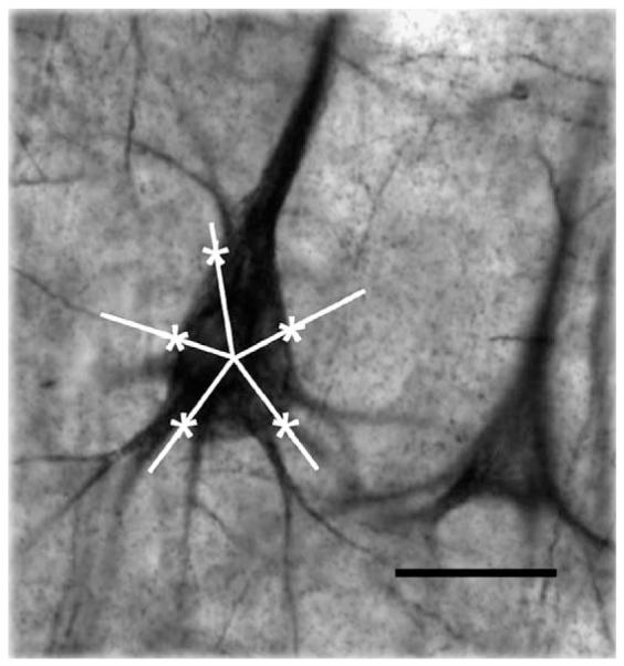
Estimation of somal size with the nucleator. The photomicrograph shows a NF200-immunoreactive neuron of area 9 with an overlay of five equally spaced rays centered within its soma. The intersections of each ray with the outline of the soma were marked to obtain an estimate of somal size using the stereological nucleator probe. Calibration bar: 25 μm.