


Figure 2.
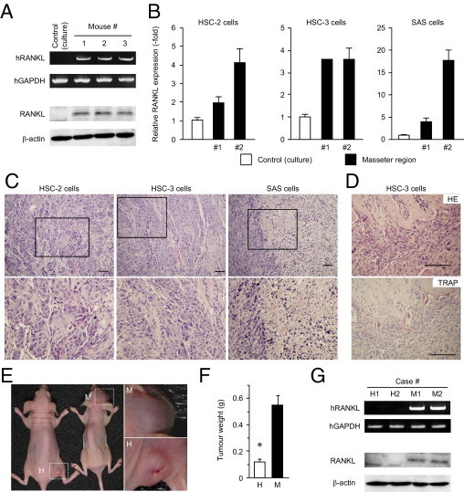
Induction of RANKL expression in the head and neck environment. A: Human head and neck cancer cells (HSC-3) were inoculated into nude mice and allowed to form tumors. Expression levels of RANKL mRNA (upper rows) and protein (lower rows) were analyzed by RT-PCR and immunoblotting, respectively. Cells under culture condition before inoculation to mice were used as a control. B: Cells of the HNSCC lines were inoculated into nude mice and allowed to form tumors. RANKL mRNA levels were evaluated by qPCR. Of the cell lines tested, HSC-2, HSC-3, and SAS cells formed tumors. Cells under culture conditions before inoculation were used as a control (open column). C and D: The tissues of tumors formed by the three cell lines were subjected to histopathological examination under H&E staining (HE) (C and D, top) and tartrate-resistant acid phosphatase staining (TRAP) (D, bottom) are shown. Scale bars = 100 μm. E–G: HSC-3 cells were injected into the masseter (M) or hindlimb (H) region of the mice. After 28 days, the tumors that formed were photographed (E) and weighed (F) (*P < 0.05). Expression levels of RANKL mRNA and protein in the masseter region (M1 and M2) or the hind leg region (H1 and H2) of nude mice were analyzed by RT-PCR (G, upper rows) and immunoblotting (G, lower rows).