


Figure 3.
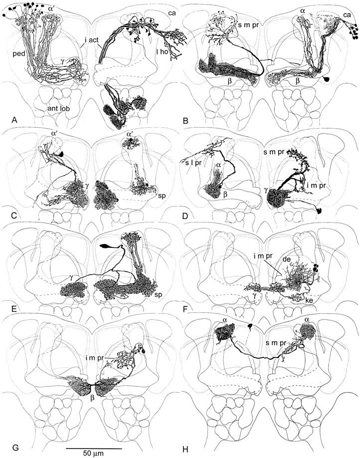
Camera lucida drawing of Golgi impregnated mushroom body neurons against neuropil outlines. (A, left) Kenyon cells with clawed dendrites in the calyx (ca) send axons through the pedunculus (ped) to γ and α′ lobes where they form whorls within terminal swellings. (A, right) Antennal lobe (ant lob) projection neurons comprise the major supply to the calyx (ca) via the inner antennocerebral tract (i act), extending into the lateral horn (l ho). (B, left) Efferent neuron from the β lobe, with terminals in α and in surrounding s m pr. (B, right) Kenyon cells with slender spined dendrites in the calyx supply the β and α lobes. (C, left) An efferent neuron originating from terminal swellings of γ sends recurrent branches back into γ and an axon to the head of α′ and surrounding s m pr. (C, right) Glial cells in γ swellings, in the ped-α′ junction near the spur (sp), and at the head of α′ occupy distinct domains corresponding to specific extrinsic neuron processes. (D, left) Efferent neuron arising from the pedunculus-α lobe junction, with terminals in the roof of the s l pr and part of the s m pr. (D, right) Efferent neuron from terminal swellings of γ, with endings in the s m pr and i m pr. (E) Protocerebral “interior” neuron supplying heterolateral arborizations to γ and unilateral branches to the head of α′. (F) Afferent extrinsic neuron with dendrites (de) in the i m pr, providing heterolateral terminals to dorsal swellings of γ (see Fig. 2E), and recurrent endings in the keel (ke) of γ. (G) Heterolateral efferent neuron originating from the terminal swelling of β and providing varicose terminals in the i m pr, flanking the α lobe shaft. (H) Heterolateral neuron linking the heads of α, with arborizations in the s m pr. Cell body location uncertain.