


Fig. 2.
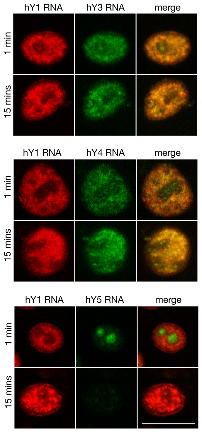
Colocalisation of human Y RNAs. Alexa-Fluor-conjugated hY RNAs were co-incubated with late G1 phase nuclei in the presence of fractions QA and ArFT for 1 minute and 15 minutes, as indicated. Nuclei were fixed and fluorescent hY RNAs were directly visualised by confocal microscopy. Single channels and merged images of Alexa-Fluor-594-conjugated hY1 RNA (red) and Alexa-Fluor-488-conjugated hY3, hY4 and hY5 RNAs (green) are shown for representative nuclei. Scale bar: 10 μm.