


Figure 2. Distribution of gene-expressing cells after i.d. DNA (gWiz-GFP) injection and non-invasive EP.
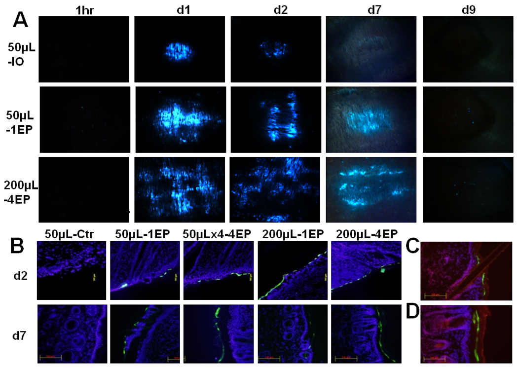
Skin samples were collected post-delivery, 1hour, day 1, day 2, day 7 or day 9. Samples were analyzed by immunoflurescence microscopy. Delivery group, 50µL-IO: 50µL DNA without EP; 50µL-1EP: 50µL DNA with 1 EP on the injection site; 50µLx4-4EP: 4 injection of 50µL DNA and each EP on the injection site; 200µL-1EP: 200µL DNA and 1 EP; 200µL-4EP: 200µL DNA and 4 EPs. A, One representative picture of 3 treated sites. (B, C, D) Total 6 cryosections (2 sections per sample) of each delivery were analyzed. Cell nuclei were blue-stained by DAPI. GFP-expressing cells were shown green. (C, D) Cell nuclei and stratum corneum was shown red-stained by propidium iodide. B, One representative section of each delivery was presented for post-delivery day 2 and day 7 (magnification = 100, scale bar=100µm). C, One representative section from post-delivery day 2 (magnification = 200). D, One representative section from post-delivery day 7 (magnification = 200, scale bar=100µm).