


Figure 4. Effects of p190RhoGAP knockdown on protective effects by OxPAPC in the in vitro model of VILI.
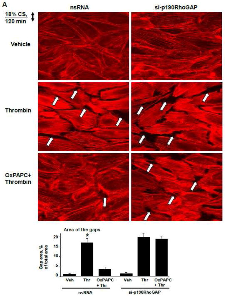
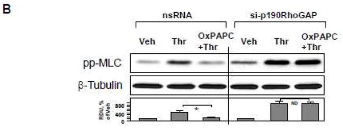
HPAEC grown on Flexcell plates were transfected with p190RhoGAP-specific siRNA or non-specific RNA duplexes. After 72 hr cell monolayers were preconditioned at pathologic (18%) levels of CS for 2 hr and stimulated with thrombin (0.1 U/ml, 15 min) with or without OxPAPC pretreatment (10 μg/ml, 15 min). A: F-actin was visualized by immunofluorescence staining with Texas-Red phalloidin. Paracellular gaps are marked by arrows. Bar graphs represent quantitative analysis of gap formation in control and treated HPAEC. Data are expressed as mean ± SD of three independent experiments; *p<0.05. B: Phosphorylation of MLC was detected by western blot with phospho-specific antibodies. Equal protein loading was confirmed by re-preobing of membranes with β-tubulin antibody. Result of densitometry shown as mean ± SD, * p<0.05