


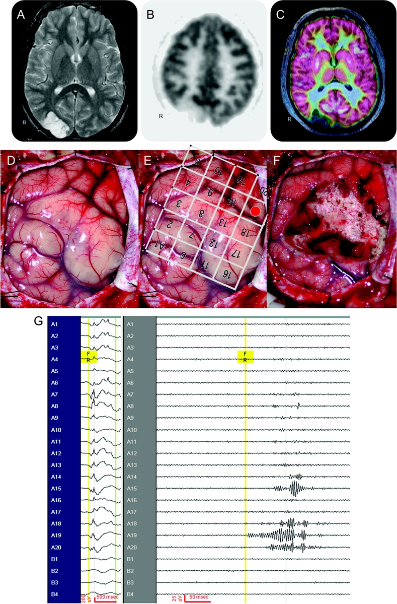
Figure 2 An illustrative case (patient 26)
This is a 16-year-old patient with 4 years of intractable epilepsy described as visual blurring and distortion. His video-EEG did not show clear electrographic onset during all stereotypical events (not shown), and his head MRI (A) showed a lesion over the right occipital region. His 18fluoro-deoxyglucose PET (FDG-PET) (B) showed hypometabolism over the right occipital region, and the coregistration of MRI and FDG-PET (C) demonstrated hypometabolism limited to the lesion. At surgery, the occipital lesion is seen in the surgical field (D). A 4 × 5 macroelectrode grid was placed over the surgical field (E), with a 1 × 4 macroelectrode strip off the surgical field (not shown) in the right frontal region as “control.” Intraoperative electrocorticography showed intermittent slowing, attenuation, and sharp waves superimposed with paroxysmal fast activity (G, left screen, no filters, no notch filter, time scale 30 mm/s, referential montage to contralateral frontal scalp electrode). After the lesion was resected (F), interictal fast ripples (FRs) (G, right screen, 240-Hz high-pass filter and 500-Hz low-pass filter, time scale 338 mm/s, referential montage to contralateral frontal scalp electrode) were seen first over A19 (E, shaded red) in the parietal region, with quick propagation to adjacent A20 and A15 superiorly, and A18 inferiorly, and importantly not found in the “control” frontal lobe. Thus the occipital lesion was resected but the parietal region containing FR remained. The patient's seizures returned in 3 months with similar semiology of visual distortion and blurring, again without electrographic changes in a second video-EEG. A second surgery 10 months after the first resection revealed similar electrocorticography, and FR was again found in the right parietal region. This FR-containing region was resected in the second surgery, and patient has been seizure-free for 20 months since the second resection, now off all antiepileptic drugs. Final pathology showed dysembryoplastic neuroepithelial tumor in the first resected specimen, and residual tumor in the second resected specimen.