


FIGURE 2.
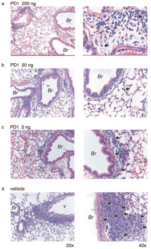
Lung histopathology from mice given PD1. Mice were sensitized and aerosol challenged with OVA in the presence of PD1 (200 ng (a), 20 ng (b), 2 ng (c)) or vehicle (d). Representative (n ≥ 3) lung tissue sections (original magnifications, ×20 (left column) and ×40 (right column)) were obtained from fixed, paraffin-embedded lung tissue, prepared, and stained with H&E. Arrows denote representative EOS; Br, bronchus; v, vessel.