


Figure 2.
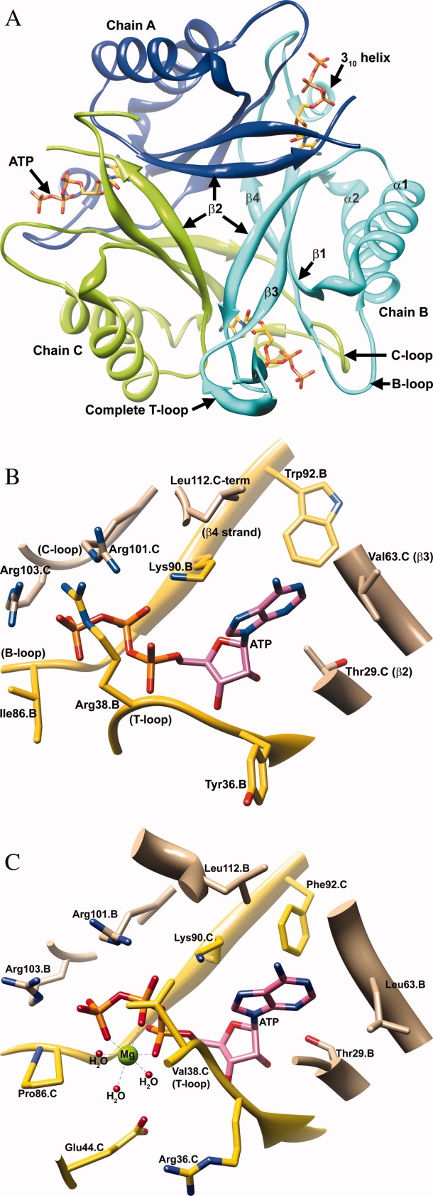
A). A trimer of the Mtb PII:ATP binary complex structure is shown viewing down the crystallographic triad. The orientation and color scheme for the trimer assembly is consistent with Figure 1A. Subunit A (blue), B (cyan), and C (green) are represented as ribbons, whereas the bound ATP molecules are shown in yellow stick representation. A complete T-loop is visible in subunit B. (B) The active site of the Mtb PII protein bound with ATP (pink). Residues contributed by the neighboring subunit are shown in tan. Residues from the main-chain (Subunit B) are shown in yellow. Conserved residues Gly87.B (B-loop), Gly89.B (B-loop), Lys90.B (β4 strand), and Arg103.C (C-loop) hydrogen bond with the polar phosphate groups of ATP. Residues contributing to adenine binding include Ile7.B (β1 strand), Trp92.B (β4 strand), Leu26.C, Gly27.C, Met28.C, and Thr29.C (in the loop connecting the α1 helix and β2 strand), Val64.C (β3 strand), and Leu112.C (C-loop). (Not all the residues in the ATP binding pocket have been displayed in the interest of clarity). (C) A side by side comparison of the ATP binding pocket of Mtb PII:ATP and the M. jannaschii GlnK1:ATP(Mg2+):2OG ternary complex structure (PDB ID: 2J9E) is presented.35 Note that the Mtb PII's Arg38 residue is a Val38 in M. jannaschii GlnK1. The side chain of Arg38 of Mtb PII occupies the Mg2+ position of M. jannaschii GlnK1.