


Figure 4. Deletion of Wip1 enhanced basal and IR-induced γ̃H2AX levels in cells undergoing oncogenic stress.
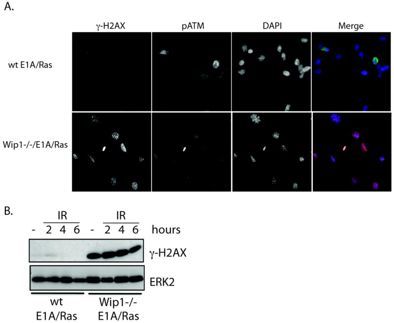
A) Immunocytochemistry of Wip1-/- E1A/Ras MEFs showed enhanced basal γ̃H2AX levels compared to wt E1A/Ras MEFs in the absence of p-ATM staining. B) Immunoblot analysis of γ-H2AX showed that Wip1-/- E1A/Ras MEFs had elevated γ-H2AX levels basally and after IR at the indicated time points compared to the wild type control (“wt E1A/Ras MEFs”). ERK2 is used as a loading control.