


Figure 4. EGFR tagged with nuclear localization sequence confers resistance to cetuximab in vitro.
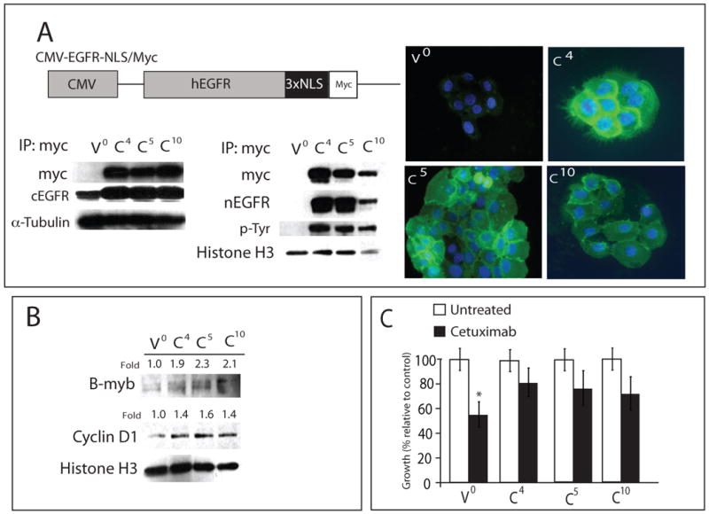
A) A schematic representation of the CMV-EGFR-NLS/Myc construct is shown. EGFR-NLS/Myc was driven by the CMV promoter. The cetuximab-sensitive NSCLC line NCI-H226 was infected with indicated constructs. Represented is three individual clones and vector control (V0; vector only, C4, C5 and C10). Cytoplasmic and nuclear protein from each clone was collected and immunoprecipitated with an anti-myc antibody, fractionated on SDS-PAGE and immunoblotted with the indicated antibodies. α-tubulin and histone H3 were used as loading and purity control for cytosolic and nuclear fractions, respectively. Immunofluorescence of nEGFR staining in CMV-EGFR-NLS/Myc clones. EGFR (green), DNA (blue), stained by PI. V0; vector clone, C4, C5 and C10; CMV-EGFR-NLS/Myc clones. 400× magnification. cEGFR; cytoplasmic EGFR, nEGFR; nuclear EGFR.
B) CMV-EGFR-NLS/Myc expressed in NCI-H226 leads to increased cyclin D1 and B-myb expression. Nuclear protein from EGFR-NLS/myc clones was collected and fractionated by SDS-PAGE followed by immunoblotting for the indicated proteins. histone H3 was used as a loading control. Expression of cyclin D1 and B-myb in CMV-EGFR-NLS/Myc clones (C4, C5 and C10) were quantitated using ImageJ software and normalized against the amounts of those proteins in vector control (V0). V0; vector clone, C4, C5 and C10; CMV-EGFR-NLS/Myc clones.
C) Growth response to cetuximab of three individual clones and vector control (V0; vector only, C4, C5 and C10). CMV-EGFR-NLS/Myc-tag clones (C4, C5 and C10) were treated with 100 nM of cetuximab and growth was measured using the growth proliferation assay and plotted as growth relative to untreated control. Data points are represented as mean +/- SEM. (n=3). *, P<0.05