


Figure 5.
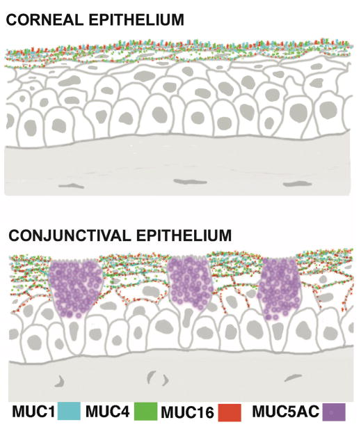
Diagram of sections of corneal and conjunctival epithelium showing the localization of expression of the membrane-spanning mucins MUC1, 4 and 16 in apical cells and the gel-forming mucin MUC5AC in goblet cells. There is very little expression of MUC4 in central epithelia. (Figure after Gipson, 2004.11)