


Figure 3.
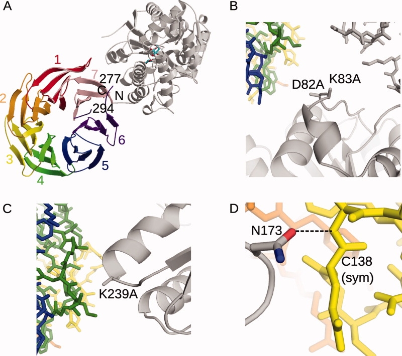
Crystal structure of the MBP(C)-RACK1A fusion protein (PDB ID code 3DM0). A: Ribbon diagram of the MBP(C)-RACK1A fusion protein. The bound maltose is drawn in stick (cyan). The structure of the MBP is shown in gray, relative to that of the RACK1A (multicolored). The β-strands of the RACK1A β-propeller domain are shown as ribbons, with each WD motif colored and labeled. The residues of the disordered loop (Lys277-Lys294) in WD6 (purple) are labeled. B,C, and D: Secondary structural elements of the MBP(C) protein are drawn as gray ribbons, with the SER mutations drawn in stick. The positions of symmetry-related MBP molecules (gray) and RACK1A (green, blue, yellow) are drawn in stick. B: Position of the D82A/K83A SER mutations in crystal lattice formation. C: Position of the K239A mutation in crystal lattice formation. D: Role of Asn173 in crystal lattice formation.