


FIGURE 9. C-terminal domain of OprF majority conformer is not exposed on cell surface.
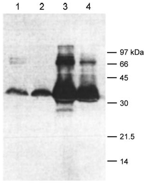
E. coli DH5α cells expressing the A312C mutant OprF were labeled with biotin-maleimide either as intact cells (lanes 1 and 2) or as crude envelope fraction after cell disruption by sonication (lanes 3 and 4). Labeling was carried out either after 2-mercaptoethanol treatment (lanes 1 and 3) or without such treatment. Fractions each containing 50 μg of protein were separated by SDS-PAGE, and the blot was stained with streptavidin reagent for biotin as described under “Experimental Procedures.”