


Figure 1.
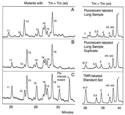
Reproducibility of the procedure and identification of mutants. Restriction-digested DNA containing the equivalent of ≈3 × 10⁸ copies of the mitochondrial target sequence was divided into two samples, and each was doped at 10−4 with two internal standards. A higher melting temperature mutant originally isolated from human tissue, mutant 13 (T → C, base pair 10,072), and a constructed mutant with a lower melting temperature than wild type (G → A, base pair 10,040) were used as internal standards. The samples were subjected to the mutational spectrometry procedure as described in Materials and Methods. (A and B) The results of CDCE separations of mutant homoduplexes isolated from duplicate DNA samples. The x axis reflects the time since the beginning of the run at which the peak reaches the detector and the y axis marks the relative intensity of fluorescence. (Left) Mutants with melting temperatures higher than wild type. (Right) Mutants with melting temperatures lower than wild type. The fluorescein-labeled samples were separated at the same time and in the same capillary as the tetramethylrhodamine (TMR)-labeled standard set containing all previously identified peaks. (C) TMR-labeled standard set separated simultaneously with the sample in B. Peaks in the samples were identified based on comigration with TMR-labeled standards and are numbered accordingly.