


Fig. 1.
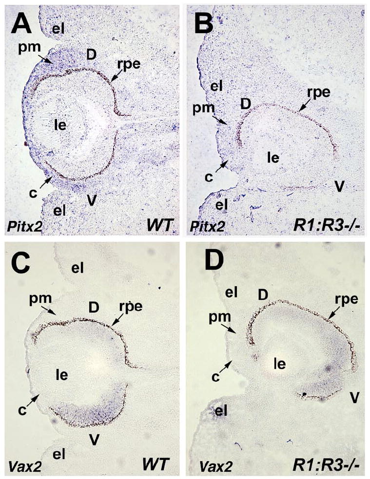
Ablation of Raldh1 and Raldh3 impairs Pitx2 expression in perioptic mesenchyme. (A-B) In situ hybridization with digoxigenin-labeled Pitx2 antisense riboprobe on frontal sections through the eye of E12.5 wild-type (WT) and double null (Raldh1-/-;Raldh3-/-) embryos. Note that Pitx2 expression in the perioptic mesenchyme of R1;R3-/- embryo is severely reduced compared to WT control indicated by arrow. (C-D) Vax2 shows normal expression pattern in the ventral retina of wild-type (WT) compared to double null (Raldh1-/-;Raldh3-/-) eyes, however note the dorsoventral rotation of the optic cup in the R1;R3-/- double mutant. c, cornea; d, dorsal retina; el, eyelid fold; le, lens; pm, perioptic mesenchyme; rpe, retinal pigment epithelium; v, ventral retina.