


Fig. 1.
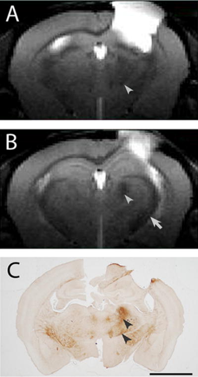
A, B) T2 weighted images, 0.5 mm thick, coronal, and centered at −1.0 mm (A) and −1.5 mm (B) relative to Bregma from an aged C57BL/6 mouse (~24 months) given a CCI injury 2 months previously. The primary injury site appears as a hyperintensity in the sensorimotor cortex. T2 hypointense areas are present in the ipsilateral thalamus (arrowheads) and ipsilateral internal capsule (arrow) relative to the contralateral structures. C) Abnormal iron deposits (arrowheads), revealed by iron histochemistry, co-localize to the T2 hypointensity in the thalamus (the same animal as for A, B). Bar = 2 mm (A–C).