


Figure 2.
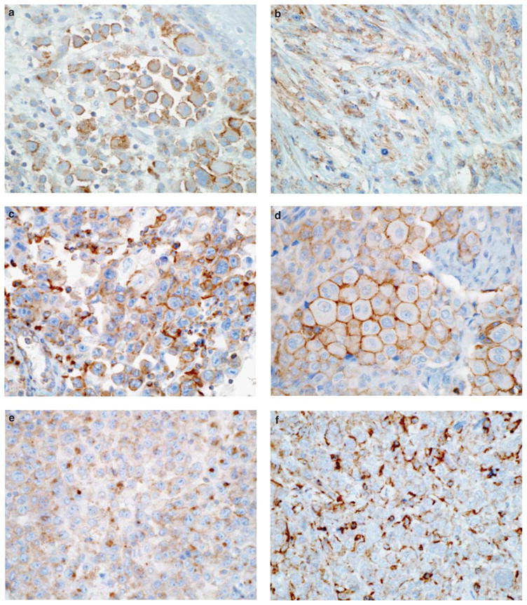
Immunohistochemical staining of cortactin in human melanomas. (a and b) Strong staining in invasive melanoma, with an accentuated cell periphery (a) and dot-like (b) distribution. (c–f) Increased expression in metastatic melanomas. The protein is most commonly seen at the cell periphery (c, d and f), with resemblance to membrane ruffles seen in cultured cells. In occasional cases, it is seen as a globule in the cytoplasm (e).