


Figure 1.
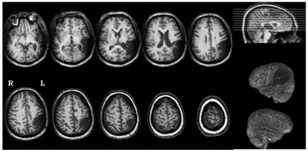
Structural T1-weighted MRI scan for patient YCR (phonologic alexia, two years poststroke). Lesion was present in cortex and subjacent white matter in temporal and parietal lobes, including Wernicke’s area, posterior middle and superior temporal gyri, angular gyrus, supramarginal gyrus, and portions of inferior and superior parietal lobules.