


Figure 4.
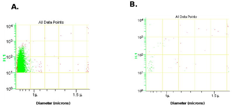
Tissue factor-bearing microparticles were measured in the conditioned media from the ASPC-1 pancreatic cancer cell line. The x-axis presents particle diameter and the y-axis presents fluorescence using a humanized anti-TF monoclonal antibody. The scatter plots for anti-tissue factor-Alexa 488 (A) and IgG-Alexa 488 control (B) are shown.