


Figure 7.
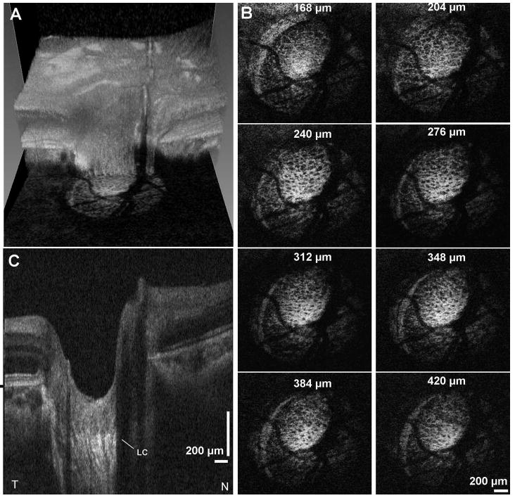
Imaging of the lamina cribrosa. (A) Images are interpolated onto en face planes through the 3D OCT data set at different depths relative to the RPE. (B) Interpolated images show the porous structure of the lamina cribrosa. Depths relative to Bruch's membrane are shown. (C) A series of six consecutive images compounded by median filtering along the slow axis shows enhanced visualization of deeper structures. The lamina cribrosa (LC) is clearly visible. Beam diameter at cornea: 1.4 mm. 512 × 450 axial scans.