


Figure 6.
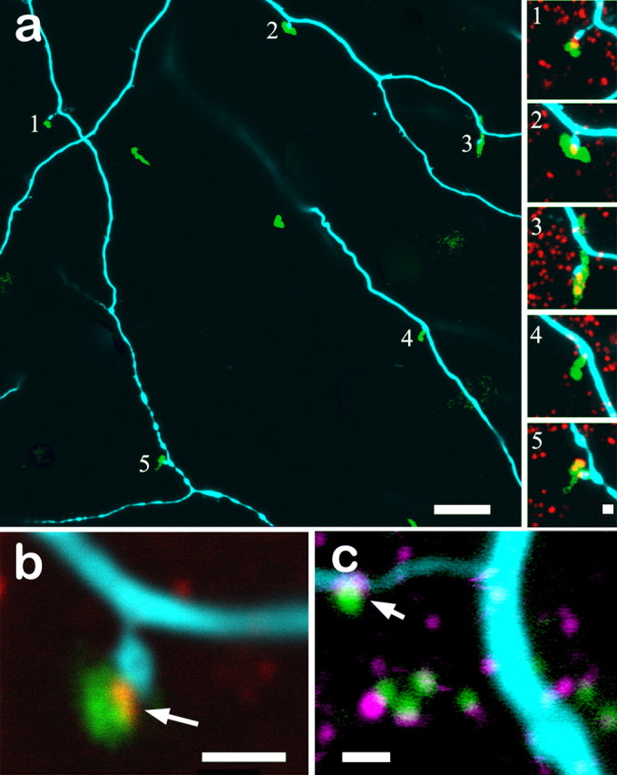
a, The dendritic arbor of the bistratified diving ganglion cell shown in Figure 5 contains numerous spines normally associated with synaptic activity in sublamina a (cyan). These spines frequently contact the descending axons of calbindin-positive bipolar cells (green). All five spines in this field also contain RIBEYE immunoreactivity (red; insets a1–a5) in which the spines contact the axons. b, Higher magnification of another spine–axon contact point clearly shows the presence of RIBEYE (red) at the conjunction of the bistratified diving ganglion cell spine (blue) and the calbindin-positive bipolar cell axon (green). c, Contact points of the bistratified diving ganglion cell (cyan) with calbindin-positive bipolar cells axons (green) also contain glutamate receptors, as seen by immunostaining of the AMPA receptor GluR4 (magenta). Scale bars: a, 10 μm; a1–a5, b, c, 1 μm. Nine optical sections of 0.3 μm thickness are stacked in a. All other panels are single 0.3 μm sections.