


Figure 2. Most CD11c+ myeloid DCs are BDCA-1− in psoriasis lesional skin.
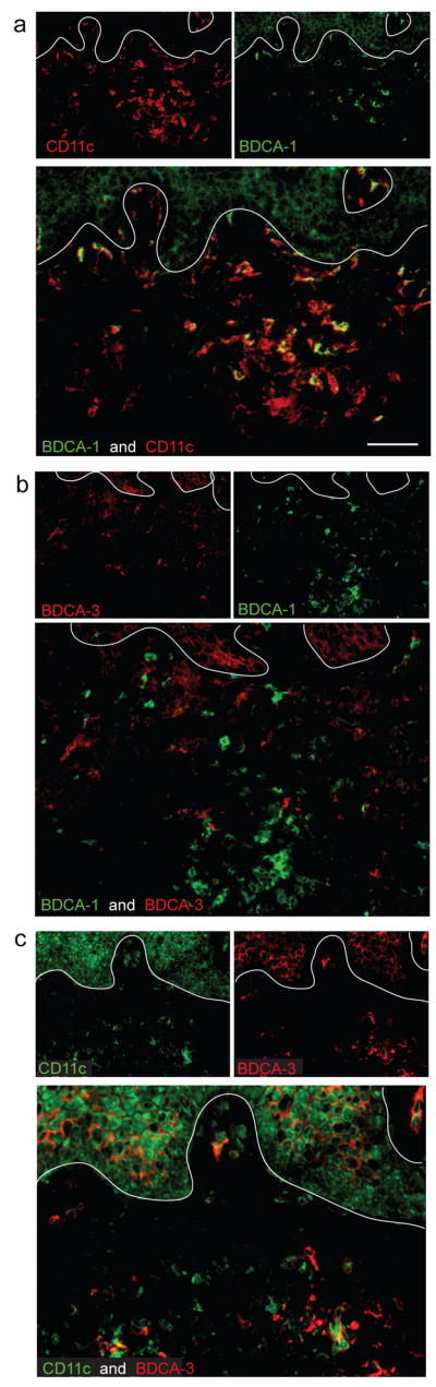
(a) The majority of CD11c+ cells in psoriatic dermis were BDCA-1−, while a small subset of CD11c+ cells co-expressed BDCA-1+. (b) BDCA-1 and BDCA-3 identified separate myeloid DC populations in the dermis. (c) Most BDCA-3+ cells co-expressed CD11c, and some BDCA-3 staining was observed on blood vessels. In all IF figures, single stained controls are above the merged image, white line denotes dermo-epidermal junction, dermal collagen fibers gave green autofluorescence, and antibodies conjugated with a fluorochrome often gave background epidermal fluorescence. Size bar = 100μm.