



Abstract
Cancer is a leading cause of mortality throughout the world and new treatments are urgently needed. Recent studies suggest that bone marrow-derived mesenchymal stem cells (MSCs) home to and incorporate within tumor tissue. We hypothesised that MSCs engineered to produce and deliver TNF-related apoptosis-inducing ligand (TRAIL), a transmembrane protein which causes selective apoptosis of tumor cells, would home to and kill cancer cells in a lung metastatic cancer model.
Human MSCs were transduced with TRAIL and the IRES-eGFP reporter gene, under the control of a tetracycline promoter using a lentiviral vector. Transduced and activated MSCs caused lung (A549), breast (MDAMB231), squamous (H357), and cervical (Hela) cancer cell apoptosis and death in co-culture experiments. Subcutaneous xenograft experiments confirmed directly delivered TRAIL-expressing MSCs were able to significantly reduce tumor growth (0.12 cm3 (0.04-0.21) vs 0.66 cm3 (0.21-1.11) (p<0.001)). We then found using a pulmonary metatastasis model, systemically delivered MSCs localised to lung metastases and the controlled local delivery of TRAIL completely cleared the metastatic disease in 38% of mice compared to 0% of controls (p<0.05).
This is the first study to demonstrate a significant reduction in metastatic tumor burden with frequent eradication of metastases using inducible TRAIL-expressing MSCs. This has a wide potential therapeutic role, which includes the treatment of both primary tumors and their metastases, possibly as an adjuvant therapy in clearing micrometastatic disease following primary tumor resection.
Keywords: Mesenchymal Stem Cell, Lung Cancer, Breast Cancer, TRAIL, Apoptosis
Introduction
Cancer remains one of the biggest causes of mortality and morbidity throughout the world (1). Present therapy focuses on varying combinations of surgery, chemotherapy and radiation treatment. Despite healthcare improvements, metastatic disease remains poorly responsive to conventional therapy and a new modality of treatment is urgently needed.
Bone marrow-derived mesenchymal stem cells (MSCs) are stromal cells that reside within the adult bone marrow. They are characteristically able to differentiate into bone, cartilage and fat, and have roles in the differentiation of haematopoietic cells. Recent studies have shown an ability of these cells to migrate to and incorporate within the connective tissue stroma of tumors (2-7). This property of MSCs can be utilised to direct targeting antitumor agents to tumor cells and their micrometastases with improvement in murine tumor models of glioma (4, 8), melanoma (6), and breast (5) and colon (7) cancers.
Tumor necrosis factor related apoptosis inducing ligand (TRAIL) is a type 2 transmembrane death ligand that causes apoptosis of target cells through the extrinsic apoptosis pathway. TRAIL is a member of the tumor necrosis factor (TNF) superfamily, which includes TNF and Fas-L (9). The expression of TNF and Fas-L leads to the damage of normal tissues in addition to their proapoptotic effect on transformed cells (10, 11), limiting their clinical applications. Conversely, TRAIL is able to selectively induce apoptosis in transformed cells, but not in most normal cells (9, 12, 13), making it a promising candidate for tumor therapy. Intravenous delivery of recombinant TRAIL has however met with problems including a short pharmacokinetic half-life (12), necessitating frequent and high doses to produce the desired effect. The use of MSCs as a delivery vector promises to provide both targeted and prolonged delivery of this death ligand.
In this study we express TRAIL in MSCs using a lentivirus conditionally activated by doxycycline. This system allows a mixed cell and gene therapy approach for metastatic cancers that can be activated and deactivated. We show that MSCs can be infected at high efficiency using the lentivirus system and delivery of trail causes apoptosis of cancer cells through the extrinsic death pathway. In vivo models confirm a prediliction of engraftment of MSCs within metastatic lung tumors with activation of TRAIL resulting in a significant reduction in metastasis number and complete clearance in 38% of mice.
Materials and Methods
Cell Culture
Tissue culture reagents were purchased from Invitrogen (Paisley, UK) unless otherwise stated. All cells were obtained from Cancer Research UK, London Research Institute (CRUK, London, UK) and were cultured in DMEM and 10% fetal bovine serum (FBS), unless otherwise stated. Human adult mesenchymal stem cells were purchased from Tulane University (New Orleans, USA) and cultured in αMEM with 16% FBS. Adipogenic and osteogenic differentiation of MSCs was performed, as previously described. (14, 15).
TRAIL lentivirus construction and transfection of MSCs
A lentiviral plasmid (pRRL-cPPT-hPGK-mcs-WPRE) into which the Tet-on system elements had been introduced (16) (a kind gift from O. Danos, UCL, London, UK), was used as a backbone for the incorporation of TRAIL DNA. The existing reporter gene, MuSEAP was excised using the Mlu1 and EcoRV restriction sites. The IRES-eGFP sequence (from pENTR1A) was amplified and restriction sites introduced by PCR and then inserted into the plasmid in place of MuSEAP. Subsequently, human TRAIL (amino acids 1-281) (RZPD, Berlin, Germany) was similarly amplified and restriction sites introduced by PCR and then cloned into the lentivirus plasmid, via Mlu1 and BstB1 restriction sites, next to the IRES-eGFP. The plasmid constructs were confirmed by DNA sequence analysis (Cogenics, Essex, UK).
The lentivirus was produced by transfecting 293T cells with 15mls of Optimem plus 10mls of a solution made by mixing 3.6ml polyethylenimine (PEI) (Sigma Aldrich) and 56.4ml Optimem, to 600μg TRAIL plasmid, 450μg of the packaging construct pCMV-dR8.74, 150μg of a plasmid producing the VSV-G envelope, pMD.G2, and 60mls Optimem (both pCMV-dR8.74 and pMD.G2 were a kind gift from A. Thrasher, UCL, London, UK). The lentivirus was concentrated by ultracentrifugation at 18000 rpm (SW28 rotor, Optima LE80K Ultracentrifuge, Beckman) at 4°C and stored at −80°C before use.
Human mesenchymal stem cells were transduced with a multiplicity of infection (MOI) of 10 virus particles for each cell and 4μg/ml of polybrene (Sigma Aldrich, Poole, UK).
Human TRAIL expression was verified by ELISA (R&D Systems, Abingdon, UK) as per manufacturer's instructions and Western blot.
Western blots
Supernatants were centrifuged, and cells lysed in RIPA buffer (PBS, 1% Igepal Ca-630, 0.5% Sodium deoxycholate, 0.1% sodium dodecyl sulphate (Sigma)) supplemented with complete protease inhibitor cocktail (Complete-mini; Roche Diagnostics Ltd, Burgess Hill, UK). Samples were denatured, resolved with a 10% SDS-polyacrylamide gel, and transferred using a semi-dry transfer method (NovaBlot; Pharmacia LKB, Sweden).
Blots were then incubated with anti-TRAIL (1μg/ml H257 rabbit polyclonal antibody (17) (Santa Cruz Biotechnology, Inc., California, USA)) or anti-actin (0.24μg/ml, Sigma) antibodies and detected using enhanced chemiluminescence (GE Healthcare, Bucks, UK) as per manufacturer's instructions.
Co-culture experiments
Human MSCs permanently transduced with the full length TRAIL plasmid (MSCFLT) were plated with target cells. A total of 100000 cells were plated in each 6-well plate. The following day, doxycycline, or other active agents were added to the cocultures and left for 48 hours. Staining of target cells was performed with the fluorescent dye, DiI, as per manufacturer's instructions (Invitrogen, Paisley, UK).
Apoptosis and cell death was assessed by flow cytometry (FACSCalibur or LSRII machines (Becton Dickenson, Oxford, UK)). This was performed on floating and adherent cells from cocultures using Annexin V-647 antibody (Invitrogen, Paisley, UK) and 5μg/ml Propidium Iodide (PI) (Sigma). DAPI (2μg/ml) (Sigma) was used instead of PI when cancer cells were stained with DiI. Annexin V−/PI− cells were judged to be non-apoptotic. Annexin V+/PI− cells were considered to be apoptotic. Annexin V+/PI+ cells were recorded as being dead.
The following compounds were used in co-culture experiments; the pancaspase inhibitor zVADfmk (1μg/ml) (Sigma), a soluble recombinant TRAIL (200ng/ml) (R & D Systems, Abingdon, UK) and a neutralising TRAIL antibody (250ng/ml) (R & D Systems).
Production of dominant negative FADD cancer cells
Phoenix packaging cells were transiently transfected with retroviral vector for the dominant negative FADD/GFP construct (dnFADD) (18) or empty vector and the virus containing supernatant was added to infect AM12 packaging cells as previously described (19). The AM12 cell supernatant was added to Hela cells (30% confluence) with 5μg/ml polybrene and the cells were selected with 1μg/ml puromycin (20).
Cell migration assay
An in vitro cell migration assay was performed to determine the tropism of MSCs for tumor cells according to previously described methods (4, 7, 21). 150000 cancer cells or 293T cells were plated in 800μl medium on the bottom well of a transwell plate (8μm pore membrane; Becton Dickenson) for 24 hours before 40000 MSCs in 300μl of the same medium were added to the upper well. The MSCs were allowed to migrate across the membrane for 24 hours at 37°C. The cells attached to the upper side of the membrane were removed, and the migrated cells on the lower side fixed, stained using a Rapid Romanowsky staining kit (Raymond Lamb, Eastbourne, UK), and counted (5 fields/well, triplicate wells) at ×10 magnification (Olympus BX40, Watford, UK).
Xenograft cancer models
All animal studies were performed in accordance with British Home Office procedural and ethical guidelines. Six-week old NOD/SCID mice (Harlan, Bicester, UK) were kept in filter cages.
Subcutaneous tumors were obtained by the injection of two million MDAMB231 cells in 200μl PBS, subcutaneously into the left flank with a 29G needle. Tumors were measured every 3-5 days with callipers, and the volume calculated as 4/3πr3, where r denoted the radius. Metastatic lung tumors were produced by the intravenous delivery of two million MDAMB231 in 200μl PBS into the lateral tail vein.
In vivo use of TRAIL-expressing mesenchymal stem cells (MSCFLTs)
In the subcutaneous models, MSCFLTs, labelled with CM-DiI (Invitrogen) as per manufacturer's instructions, were either delivered concurrently with the cancer cells; 2 million MDAMB231 with 0.75 million MSCFLT in 200μl PBS, or after tumors had become established; 1 million MSCFLTs were injected into established subcutaneous tumors. In metastatic models, 0.75 million MSCFLT cells were suspended in 200μl PBS and injected into the lateral tail vein at 7, 14, 21 and 28 days after the cancer models had been set up (5). To activate the TRAIL constructs in the transduced MSCFLTs, mice were fed water containing 2mg/ml doxycycline and 3% sucrose, with controls fed water with 3% sucrose only.
Mice were sacrificed by CO2 asphyxiation followed by exsanguination. Subcutaneous tumors were removed and weighed before they were split for digestion with 0.1% collagenase A (Roche Diagnostics Ltd., Burgess Hill, UK) into a single cell suspension as per manufacturer's protocol, or fixed in 4% paraformaldehyde for histology. The lungs were excised and weighed before they were insufflated with a fixed 20 cm pressure of 4% paraformaldehyde and then bathed in 4% paraformaldehyde for histology. In some lungs, the left lobe was removed and snap frozen in liquid nitrogen before insufflation.
Immunohistochemistry
Fixed specimens were embedded in paraffin and cut into 3μm sections for Haematoxylin and Eosin (H&E) staining. GFP antibody (Rabbit polyclonal: Invitrogen) and TRAIL antibody (Rabbit monoclonal: Santa Cruz, Heidelberg, Germany) were used as primary antibodies and detected with biotinylated secondaries and diaminobenzidine (DAB; Vector Laboratories, California, USA). Fluorescent microscopy was used to detect DiI positive cells with DAPI counterstain, and secondary fluorescent antibodies against primary vimentin antibodies (sc7557, Santa Cruz). TUNEL staining was performed according to the manufacturer's instructions (Roche Diagnostics, Burgess Hill, UK). Microscopy was performed using light (Olympus BX40, Watford, UK), fluorescent (Carl Zeiss Ltd., Axioskop 2, Welwyn Garden City, UK) or confocal microscopes (Bio-Rad MRC 1024, Hercules, California, USA).
Quantitative Real-Time RTPCR
RNA was extracted from the snap frozen left lung with TRIzol (Invitrogen) and treated with DNAse (Ambion, Texas, USA). cDNA was synthesized using a RT-PCR kit, according to manufacturer's instructions (Roche Diagnostics). Real-time PCR amplification was performed with Light Thermocycler (Roche Diagnostics) using SYBR Green qPCR kit (Invitrogen) with primers for human TRAIL and the control 18s.
Metastases Quantification
The number and size of tumor nodules were assessed in H&E sections using similar methodology to that previously described (22). Photomicrographs of representative sections of the entire lung were taken at ×4 magnification. This created a complete picture of all lobes of the lungs of the mice. Image analysis software (SimplePCI High Performance Imaging Software, Hamamatsu Photonics, Herrsching, Germany) was used to trace around the metastatic deposits and the lung sections and then calculate lung and metastasis area and number of metastases.
Statistics
Statistical analysis was performed using GraphPad Prism v4 (GraphPad Software, California, USA). Multiple groups were analysed by Anova or Kruskal-Wallis tests. Single group data was assessed using Students t-test. All in vitro experiments were performed in triplicate unless specified.
Results
Characterisation and TRAIL transduction of Mesenchymal stem cells
Fully characterised MSCs were purchased from Tulane University and were shown to differentiate into fat and bone in differentiation assays and have the expected colony forming efficiency (48% ± 2.83 (SD)) (Figure 1A). Mesenchymal stem cells were stably transduced with our lentivirus expressing TRAIL and GFP under the control of doxycycline at different multiplicities of infection (MOI). An MOI of 10 was used for further experiments producing 82.13% ± 0.40 successful transduction (GFP expression) at day seven (Figure 1B), while limiting the number of dead cells 4.8% ± 1.83).
Figure 1. MSC characterisation, transduction and gene expression.

(A) MSCs, differentiation to adipocytes (Oil-Red-O) and osteoblasts (Alizarin Red S) (×4 mag), and colony forming ability. (B) Representative flow cytometry for GFP expression in MSCs transduced with the TRAIL-GFP lentivirus (MSCFLTs) and activated with doxycycline, and timescale of GFP expression with addition or removal of doxycycline. (C) TRAIL ELISA of MSCFLTs. (D) Western blots of MSCFLTs showing maximum expression of TRAIL after two days of doxycycline treatment (10μg protein loaded) and loss of expression one day after doxycycline removal (5μg protein loaded) with actin loading controls.
MSCFLTs were examined for TRAIL and GFP expression using flow cytometry, Western blots and ELISA. Flow cytometry demonstrated <0.5% GFP expression before activation with doxycycline while 48 hours after doxycycline addition to the media, this increased to 74.7% ± 2.5 (Figure 1B). Withdrawl of doxycycline, after the cells had been exposed for five days, led to a fall in GFP expression with 12.2% ± 1.0 of cells weakly positive at day seven (Figure 1B and Supplementary Figure S1).
ELISA and immunoblotting for TRAIL demonstrated it was only produced when the transgene was activated by doxycycline (Figure 1C,D). Western blots demonstrated TRAIL protein expression in MSCFLT lysates was maximal after two days of doxycycline stimulus (Figure 1D), but very little TRAIL protein remained after the doxycycline stimulus had been removed for one day, following a preceding doxycycline stimulus of five days (Figure 1D).
Trail-expressing MSCs cause tumor cell death in vitro
MSCFLTs were co-cultured with tumor cells, and apoptotic and dead cells increased significantly when doxycycline was added. This effect was observed with lung cancer (A549) (apoptotic and dead cells increased from 3.1% ± 0.1 to 19.6% ± 0.8 (p=0.001, t-test) with the addition of doxycycline), breast cancer (MDAMB231) (15.4% ± 1.7 to 37.7% ± 6.5)(p=0.001, t-test), squamous cell cancer (H357) (12.5% ± 0.3 to 34.5% ± 1.0)(p=0.001, t-test) and cervical cancer (Hela) (6.7% ± 1.0 to 36.1% ± 3.5)(p=0.0001, t-test) cells (Figure 2A-C). These apoptosed and dead cells were shown to come specifically from the cancer cell population by labelling the cancer cell populations with the fluorescent dye (DiI). 50.7% ± 2.7 of the Hela cell population were dead or apoptotic compared to only 8.3% ± 4.6 of the MSCFLT population (p<0.0001, t-test) (Figure 2D).
Figure 2. TRAIL-expressing MSCs cause cancer cell apoptosis in vitro.
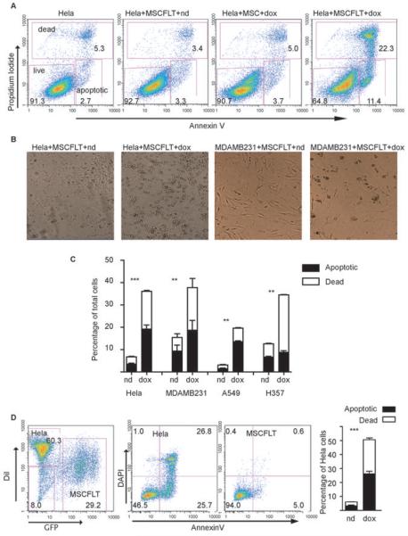
(A) Representative flow cytometry plots demonstrating an increase in death and apoptosis when Hela cells were co-cultured with doxycycline (dox) treated MSCFLTs compared to no doxycycline (nd), untreated controls or doxycycline treated normal MSCs. (B) Phase microscopy (×5 mag) demonstrating an increase in cell death (rounded and floating cells) when MSCFLTs were activated by doxycycline in co-culture with Hela and MDAMB231 cells. (C) Flow cytometry results from triplicate apoptosis assays showing an increase in death and apoptosis of total cells in cancer cell and MSCFLT co-cultures after doxycycline treatment. (D) Flow cytometry of co-culture experiments demonstrated DiI positive cancer cells were responsible for dead and apoptotic populations. Triplicate experiments shown on bar graph. (***p<0.001 **p<0.01).
Co-culture experiments were repeated with decreasing numbers of MSCFLTs. A significant increase in apoptosis and death of the cancer cells was achieved at all concentrations of MSCFLT used (p<0.001 at ratio MSCFLT:Hela cells of 1:16, Anova). There was a cell ratio-dependent effect (Figure 3A). This experiment under-estimates the true killing capacity of the MSCFLT cells as the seeding ratios do not account for any proliferation of cancer cells compared to the MSCFLTs before the addition of doxycycline.
Figure 3. TRAIL-expressing MSC-induced apoptosis occurs at low MSCFLT to cancer cell ratios via the extrinsic apoptotic pathway.

(A) Results from flow cytometry apoptosis assays showing an increase in death and apoptosis of Hela cells when MSCFLTs activated with doxycycline (dox) were co-cultured with Hela cells even at low 1:16 ratios, compared to untreated controls (nd). (B) Induced cell death and apoptosis is higher using the MSCFLTs than with recombinant TRAIL (rhTRL) and can be partially blocked with blocking antibody (Ab). (C) Cell death and apoptosis is reduced when using Hela cells expressing dominant negative Fas-associated death domain (dnFADD) in comparison with those transduced with an empty vector in addition to when zVADfmk (zvad), a pan-caspase inhibitor, is used compared to the control (con). (***p<0.001 **p<0.01 *p<0.05).
Trail-expressing MSCs kill cancer cells by TRAIL induction of the extrinsic apoptosis pathway
A TRAIL antibody with some ability to neutralise the bioactivity of TRAIL was added to the MSCFLT and Hela cell co-cultures. The antibody was used at a dose to give maximal neutralising effect (250ng/ml) (23). The amount of MSCFLT-induced death and apoptosis was significantly reduced with this antibody (38.03% ± 0.49 compared to 50.67% ± 3.8)( p<0.001, Anova) however not back to baseline (11.48% ± 1.49) (Figure 3B). We found our doxycycline-induced MSCFLTs were potent inducers of death and apoptosis of Hela cells, with a larger proportion of apoptotic and dead cells in comparison to maximal doses (200ng/ml) of recombinant soluble TRAIL (50.67% ± 3.8 compared to 27.50% ± 0.70)(p<0.001, Anova) (Figure 3B).
The caspases are a family of closely related enzymes crucial to apoptosis. zVADfmk is a cell permeable, pan-caspase inhibitor. Application of this compound to the 1:1 co-cultured MSCFLTs and Hela cells caused a 51.6% reduction in death and apoptosis (26.5% ± 0.7 compared to a DMSO treated control 54.8% ± 3.0)( p=0.003, t-test) confirming the importance of caspases in the mechanism of TRAIL-induced cell death (Figure 3C).
To confirm the mechanism of MSCFLT induced cancer cell death is via the extrinsic pathway, Hela cancer cells were retrovirally transduced with a dominant negative Fas-associated death domain (dnFADD) construct or an empty vector (18). The dnFADD consists of the death domain that binds to the TRAIL receptor, but not the domain responsible for caspase 8 recruitment and the triggering of apoptosis. We observed a significant reduction (p=0.018, t-test) in death and apoptosis of Hela cells transduced with dnFADD after co-culture with activated MSCFLT cells (Figure 3C). This data presents strong evidence the MSCFLT cells induce apoptosis via the extrinsic pathway.
MSCs migrate towards some tumor cells
In vitro transwell experiments demonstrated the ability of MSCs to home towards tumors. A549, MDAMB231, and Hela cancer cell cultures were used to attract MSCs, with benign fibroblast 293T cells and medium alone used as controls. There was significantly increased migration to the MDAMB231 cells; 250.4 ± 0.8 cells/field compared to 98.6 ± 9.3 cells to 293T and 57.6 ± 7.6 towards the medium (p<0.001, Anova). There was not a significantly increased migration towards the A549 (92.0 ± 7.1) and Hela (81.0 ± 2.1) cells (Figure 4A).
Figure 4. MSCs migrate to some cancer cells in vitro and preferentially engraft in vivo.

(A) Transwell migration studies demonstrated an increased number (per microscopic field) of MSCs migrating through the transwell membrane toward MDAMB231 cells, but not towards other (A549, Hela) cancer cell types compared to control 293T cells or medium alone (***p<0.001). (B) H&Es showing representative lung metastases post intravenous injection of MDAMB231 cells into NOD/SCID mice at day 10, 20, and 30 and a macroscopic picture at day 30. Scale bar represents 20μm. (C) DiI-labelled MSCFLTs (red) injected intravenously at day 10 and shown to localise to lung metastases on fluorescent microscopy with DAPI nuclear counterstain with H&E contiguous sections from day 30 harvested lungs ( ×10 mag scale bar represents 20μm and ×4 mag scale bar represents 60μm).
To examine if MSCFLTs could be traced to metastatic tumors we used a lung metastasis model. Intravenously injected MDAMB231 cells produced lung metastases in all of 20 NOD/SCID mice. These tumors were visible at day ten post-injection and grew to large metastases by day 30 (Figure 4B). DiI-stained MSCFLTs were injected at day 10 and shown histologically to preferentially localise to lung metastases (Figure 4C). DiI-stained fibroblasts were not detected in the tumors or indeed the lungs ten days after injection (data not shown). This histological data shows MSCFLTs preferentially engraft and are maintained in the in vivo tumor environment compared to surrounding lung parenchyma while fibroblasts are not.
Trail-expressing MSCs reduce subcutaneous tumor growth in mice
Two million MDAMB231 cells were coinjected with 0.75 million MSCFLT cells and the TRAIL transgene activated with the addition of doxycycline at day 0, day 25, or never, with tumors harvested at day 42. A recent study has demonstrated that coculture of MSCs with MDAMB231 cells did not alter the tumour growth (24). In our study with transduced MSCs, activation at day 0 resulted in a significantly reduced tumor size (0.12 cm3 ± 0.12 vs 0.66 cm3 ± 0.49)(p<0.001, 2-way Anova) and weight (0.07 g ± 0.06 vs 0.33 g ± 0.33) (p<0.05, Kruskal-Wallis), however there was no change in the tumor growth when TRAIL was activated after the tumor had already become established at day 25 (Figure 5A,B). TUNEL staining of excised tumors demonstrated areas of apoptosis co-localised with DiI-labelled MSCFLT cells (Figure 5C). The subcutaneous tumors were excised and digested to a single cell suspension. Confocal microscopy of the ex vivo DiI-labelled cells included cells of MSC type morphology and demonstrated vimentin staining (Figure 5D). A separate experiment injected transduced MSCFLTs into established tumors. Similar to late activation of co-injected cells, the late injection of MSCFLT cells into established tumors and activation of TRAIL at this later stage was unable to alter the growth of the tumors (Supplementary Figure S2).
Figure 5. TRAIL-expressing MSCs reduce the growth of subcutaneous tumors.

(A) Doxycycline (dox) treatment of subcutaneous tumors composed of mixed MDAMB231 cells and MSCFLT cells from day 0 led to a decreased size and weight (*p<0.05, **p<0.01, ***p<0.001) of the tumors compared to no doxycycline treated mice (nd). There was no reduction in tumor growth if TRAIL was activated at day 25. (B) Macroscopic appearance of the tumors. (C) TUNEL staining (green) demonstrated areas of tumor apoptosis localised with areas of MSCFLTs (DiI - red) within the tumors in mice treated with doxycycline from day 0 (DAPI nuclear counterstain - blue). Scale bar represents 25μm (D) Ex vivo single cell digestion and culture showed DiI positive (red) cells with MSC morphology that co-stained with vimentin (green) (DAPI – blue). Scale bar represents 3μm.
Systemic delivery of TRAIL-expressing MSCs reduces and can eliminate lung metastases
Intravenously injected MDAMB231 cells were used to produce lung metastases in NOD/SCID mice. MSCFLT were then used as an intravenous combined cellular and gene therapy with delivery at day 7, 14, 21 and 28 (Figure 6A). At day 35, tumor metastases were found in all mice (8/8) without MSCFLT and all mice with MSCFLT without the use of doxycycline (8/8). In the MSCFLT plus doxycycline arm, three out of eight mice were tumor free (p=0.032, Chi2) (Figure 6B). In addition to the elimination of metastases with MSCFLT plus doxycycline, the lung weight (p<0.01, Anova), which serves as a correlate for metastases load, was also significantly reduced in all of these mice. Similar results were achieved when metastases numbers per lung area was used as an endpoint (p<0.001, Anova) (Figure 6C). Three mice had to be excluded from this latter analysis as the lungs did not inflate during the fixation procedure. MSCFLT cells delivered to control mice without doxycycline were unable to clear the metastases in any mice, but did have less metastases per area (p<0.001, Anova) and a lower lung weight (p<0.05, Anova) than the untreated mice. Doxycycline treatment however produced a further significant reduction (p<0.05, Anova) (Figure 6B,C). In a subsequent experiment, TRAIL and GFP were both shown to be expressed in vivo by the MSCFLT with immunohistochemistry when mice were harvested two days after treatment with MSCFLT and doxycycline (Figure 6D) (primary antibody controls showed no staining, data not shown). TRAIL mRNA derived from the lungs of these mice was also increased (Figure 6D).
Figure 6. TRAIL expressing MSCs reduce the growth of lung metastases.

(A) 2 million MDAMB231 cells were injected intravenously at day 0 followed by delivery or not of MSCFLT cells at days 7, 14, 21 and 28 with or without doxycycline (dox – doxycycline; nd – no dox). (B) Representative histology of lung lobes in the three experimental groups. Metastases remained, but were reduced, after injection of MSCFLT without activation of the TRAIL construct, while TRAIL activation of MSCFLTs eliminated metastatses in 3 of 8 mice (p=0.03). (C) Reduction in lung weight and metastases number per lung area with the use of MSCFLTs both with and without doxycycline treatment. There was a further significant reduction between activated MSCFLTs compared to inactivated (***p<0.001 **p<0.01 *p<0.05). (D) Immunohistochemistry demonstrating cells expressing GFP (scale bar represents 5μm) and TRAIL (scale bar represents 10μm) and consecutive immunofluorescence sections showing these cells also express DiI. There is an increase in TRAIL mRNA from lung digests treated with MSCFLT and doxycycline (*p<0.05).
Discussion
The ability of TRAIL to lead to tumor apoptosis and death without affecting normal cells makes it an extremely exciting molecule for tumor therapy. Here we have shown that mesenchymal stem cells can be engineered to express TRAIL under the sensitive control of the Tetracycline-on inducible system. These cells were able to kill cancer cell lines in vitro via the extrinsic death pathway to a higher degree than recombinant protein. In vivo, we demonstrate that TRAIL-expressing MSCs reduce the growth of early subcutaneous tumors, while in a systemically delivered metastasis model these cells reduced metastases but most significantly eliminated metastases in three mice.
We used mesenchymal stem cells as vectors of delivery due to observations that they appear to home to, or at least engraft preferentially, within tumors. MSCs also make an attractive therapeutic tool as they are widely acknowledged to be immunoprivileged. Theoretically, future cell therapies using allogenic MSCs could be used in patients without the use of prior immunomodulation (25). Alternatively, with the ease of harvest, culture and infection of MSCs, the use of autologous cells may also be realistic.
It is important to consider the possible direct effects that MSCs may have on disease. Some studies have suggested intrinsic antineoplastic properties of these cells, with improvements in Kaposis's sarcoma (2) and subcutaneous breast tumor models (26). Various mechanisms have been proposed for these effects including Akt inhibition (2), NFκB downregulation (27), and the Wnt pathway (26). Conversely, mesenchymal stem cells have also been associated with a tumor promoting effect in certain models, including increased growth and metastasis in colonic (28) and breast (24) subcutaneous tumor models. In our subcutaneous tumor model we did not see an increase in lung metastases when the tumour cells were coinjected with MSCs. Furthermore, the MSCs in our lung metastasis model appeared to have an antineoplastic effect.
To our knowledge this is the first study that uses MSCs as a vector for a lentiviral delivery of a gene therapy for cancer. We used a lentivirus in view of their significant advantages over other vector systems. Adenoviral vector expression is transient and often produces a significant host immune response, wheresas retroviruses may cause incorporation errors. Lentiviral vectors are less likely to cause insertional mutagenesis as the promoter can be modified extensively. Furthermore, they have the ability to stably transduce both dividing and quiescent cells (29), which is a further significant advantage when using stem cells that are often quiescent or slow growing. Finally our construct incorporates the Tetracycline-on system allowing us to both turn on and, through withdrawl of doxycycline, off the protein expression; attractive in the view of possible longevity of using stem cells as delivery vehicles in disease. Clinically, treatment could be timed to chemotherapy or radiotherapy regimes, with the expression turned off in between courses.
TRAIL is thought to operate physiologically, in the immune surveillance against tumors. Administration of a neutralising anti-TRAIL antibody or the use of TRAIL-knockout mice demonstrated the increased susceptibility of TRAIL deficient mice to TRAIL-sensitive tumors and metastases (30-32). TRAIL-expressing MSCs in this study may act in a similar way, replacing the deficient immunosurveillance systems in the NOD/SCID mice leading to tumor cell destruction and prevention of metastasis. The property of metastasis prevention would be ideal as an adjuvant therapy in the treatment of many patients with solid organ tumors, whom remain at significant risk of future metastatic disease despite primary tumor resection and chemo and radiotherapy. Identifying these patients is now becoming a reality with the use of highly sensitive and specific molecular and cytological techniques, which allow the detection of very small numbers of circulating tumor cells in the blood and bone marrow (33, 34) which could be amenable to MSC directed TRAIL therapy.
The use of targeted TRAIL therapy could be widely applicable to many different cancers. Despite some cancers being resistant to the effects of TRAIL, an additive effect has been demonstrated with the concomitant use of TRAIL with other antineoplastic agents that act at different positions in either the intrinsic or extrinsic apoptotic pathways. For example, the downregulation of Bcl-2 (35) or the DNA damage caused by chemotherapy can increase the effects of TRAIL therapy in various in vivo models (36-39).
One of the limitations of our study is the model used for the in vivo experiments. With the aim of future translational therapeutics, we utilised human cancer xenograft models, human MSCs and human TRAIL constructs. It is important to consider that human TRAIL has only 65% homology to murine TRAIL (9), and there are also differences in the receptors in mice and humans. In the mouse, only one death-inducing receptor with homology to DR5 has been discovered, in addition to two decoy receptors (mDcTrailr1, msDcTrailr2) (40). Consequently, the effects of TRAIL on the normal murine cells may be different to the response of normal human cells.
In summary, we describe for the first time to our knowledge, the use of TRAIL expressing MSCs for the reduction and in some cases elimination of metastatic disease in a murine lung metastasis model. We believe that this therapy may have an important future therapeutic role in preventing metastatic recurrence in patients.
Supplementary Material
Acknowledgements
The authors would like to acknowledge Dr. Dominique Bonnet, Dr. Susana Aguilar, Professor Geoff Laurent and Dr. Rachel Chambers for their helpful comments on experimental design and Dr. Laura Bazley for technical assistance.
Funding: MRL is a Medical Research Council (MRC) UK Clinical Training Fellow. SMJ is a MRC Clinician Scientist. This work was partly undertaken at UCLH/UCL, which received a proportion of funding from the Department of Health's NIHR Biomedical Research Centres funding scheme.
Footnotes
Competing Interests: The authors have no interests to declare.
References
- 1.Jemal A, Siegel R, Ward E, Murray T, Xu J, Thun MJ. Cancer statistics, 2007. CA Cancer J Clin. 2007;57:43–66. doi: 10.3322/canjclin.57.1.43. [DOI] [PubMed] [Google Scholar]
- 2.Khakoo AY, Pati S, Anderson SA, et al. Human mesenchymal stem cells exert potent antitumorigenic effects in a model of Kaposi's sarcoma. J Exp Med. 2006;203:1235–47. doi: 10.1084/jem.20051921. [DOI] [PMC free article] [PubMed] [Google Scholar]
- 3.Menon LG, Picinich S, Koneru R, et al. Differential gene expression associated with migration of mesenchymal stem cells to conditioned medium from tumor cells or bone marrow cells. Stem Cells. 2007;25:520–8. doi: 10.1634/stemcells.2006-0257. [DOI] [PubMed] [Google Scholar]
- 4.Nakamizo A, Marini F, Amano T, et al. Human bone marrow-derived mesenchymal stem cells in the treatment of gliomas. Cancer Res. 2005;65:3307–18. doi: 10.1158/0008-5472.CAN-04-1874. [DOI] [PubMed] [Google Scholar]
- 5.Studeny M, Marini FC, Dembinski JL, et al. Mesenchymal stem cells: potential precursors for tumor stroma and targeted-delivery vehicles for anticancer agents. J Natl Cancer Inst. 2004;96:1593–603. doi: 10.1093/jnci/djh299. [DOI] [PubMed] [Google Scholar]
- 6.Studeny M, Marini FC, Champlin RE, Zompetta C, Fidler IJ, Andreeff M. Bone marrow-derived mesenchymal stem cells as vehicles for interferon-beta delivery into tumors. Cancer Res. 2002;62:3603–8. [PubMed] [Google Scholar]
- 7.Xin H, Kanehira M, Mizuguchi H, et al. Targeted delivery of CX3CL1 to multiple lung tumors by mesenchymal stem cells. Stem Cells. 2007;25:1618–26. doi: 10.1634/stemcells.2006-0461. [DOI] [PubMed] [Google Scholar]
- 8.Kim SM, Lim JY, Park SI, et al. Gene Therapy Using TRAIL-Secreting Human Umbilical Cord Blood-Derived Mesenchymal Stem Cells against Intracranial Glioma. Cancer Res. 2008;68:9614–23. doi: 10.1158/0008-5472.CAN-08-0451. [DOI] [PubMed] [Google Scholar]
- 9.Wiley SR, Schooley K, Smolak PJ, et al. Identification and characterization of a new member of the TNF family that induces apoptosis. Immunity. 1995;3:673–82. doi: 10.1016/1074-7613(95)90057-8. [DOI] [PubMed] [Google Scholar]
- 10.Ogasawara J, Watanabe-Fukunaga R, Adachi M, et al. Lethal effect of the anti-Fas antibody in mice. Nature. 1993;364:806–9. doi: 10.1038/364806a0. [DOI] [PubMed] [Google Scholar]
- 11.Nagata S. Apoptosis by death factor. Cell. 1997;88:355–65. doi: 10.1016/s0092-8674(00)81874-7. [DOI] [PubMed] [Google Scholar]
- 12.Ashkenazi A, Pai RC, Fong S, et al. Safety and antitumor activity of recombinant soluble Apo2 ligand. J Clin Invest. 1999;104:155–62. doi: 10.1172/JCI6926. [DOI] [PMC free article] [PubMed] [Google Scholar]
- 13.Walczak H, Miller RE, Ariail K, et al. Tumoricidal activity of tumor necrosis factor-related apoptosis-inducing ligand in vivo. Nat Med. 1999;5:157–63. doi: 10.1038/5517. [DOI] [PubMed] [Google Scholar]
- 14.Aguilar S, Nye E, Chan J, et al. Murine but not human mesenchymal stem cells generate osteosarcoma-like lesions in the lung. Stem Cells. 2007;25:1586–94. doi: 10.1634/stemcells.2006-0762. [DOI] [PubMed] [Google Scholar]
- 15.Chan J, O'Donoghue K, de la Fuente J, et al. Human fetal mesenchymal stem cells as vehicles for gene delivery. Stem Cells. 2005;23:93–102. doi: 10.1634/stemcells.2004-0138. [DOI] [PubMed] [Google Scholar]
- 16.Barde I, Zanta-Boussif MA, Paisant S, et al. Efficient control of gene expression in the hematopoietic system using a single Tet-on inducible lentiviral vector. Mol Ther. 2006;13:382–90. doi: 10.1016/j.ymthe.2005.09.012. [DOI] [PubMed] [Google Scholar]
- 17.Kagawa S, He C, Gu J, et al. Antitumor activity and bystander effects of the tumor necrosis factor-related apoptosis-inducing ligand (TRAIL) gene. Cancer Res. 2001;61:3330–8. [PubMed] [Google Scholar]
- 18.Janes SM, Watt FM. Switch from {alpha}v{beta}5 to {alpha}v{beta}6 integrin expression protects squamous cell carcinomas from anoikis. J Cell Biol. 2004;166:419–31. doi: 10.1083/jcb.200312074. [DOI] [PMC free article] [PubMed] [Google Scholar]
- 19.Janes SM, Ofstad TA, Campbell DH, Watt FM, Prowse DM. Transient activation of FOXN1 in keratinocytes induces a transcriptional programme that promotes terminal differentiation: contrasting roles of FOXN1 and Akt. J Cell Sci. 2004;117:4157–68. doi: 10.1242/jcs.01302. [DOI] [PubMed] [Google Scholar]
- 20.Legg J, Jensen UB, Broad S, Leigh I, Watt FM. Role of melanoma chondroitin sulphate proteoglycan in patterning stem cells in human interfollicular epidermis. Development. 2003;130:6049–63. doi: 10.1242/dev.00837. [DOI] [PubMed] [Google Scholar]
- 21.Abayasiriwardana KS, Barbone D, Kim KU, et al. Malignant mesothelioma cells are rapidly sensitized to TRAIL-induced apoptosis by low-dose anisomycin via Bim. Mol Cancer Ther. 2007;6:2766–76. doi: 10.1158/1535-7163.MCT-07-0278. [DOI] [PubMed] [Google Scholar]
- 22.Zhang X, Zhao P, Kennedy C, et al. Treatment of pulmonary metastatic tumors in mice using lentiviral vector-engineered stem cells. Cancer Gene Ther. 2008;15:73–84. doi: 10.1038/sj.cgt.7701108. [DOI] [PMC free article] [PubMed] [Google Scholar]
- 23.Matthews N, Neale ML. Cytotoxicity assays for tumour necrosis factor and lymphotoxin. In: Clemens MJ, Morris AG, Gearing AJH, editors. Lymphokines and interferons, a practical approach. Oxford: IRL Press; 1987. p. 221. [Google Scholar]
- 24.Karnoub AE, Dash AB, Vo AP, et al. Mesenchymal stem cells within tumour stroma promote breast cancer metastasis. Nature. 2007;449:557–63. doi: 10.1038/nature06188. [DOI] [PubMed] [Google Scholar]
- 25.Chen XC, Wang R, Zhao X, et al. Prophylaxis against carcinogenesis in three kinds of unestablished tumor models via IL12-gene-engineered MSCs. Carcinogenesis. 2006;27:2434–41. doi: 10.1093/carcin/bgl069. [DOI] [PubMed] [Google Scholar]
- 26.Qiao L, Xu ZL, Zhao TJ, Ye LH, Zhang XD. Dkk-1 secreted by mesenchymal stem cells inhibits growth of breast cancer cells via depression of Wnt signalling. Cancer Lett. 2008;269:67–77. doi: 10.1016/j.canlet.2008.04.032. [DOI] [PubMed] [Google Scholar]
- 27.Qiao L, Zhao TJ, Wang FZ, Shan CL, Ye LH, Zhang XD. NF-kappaB downregulation may be involved the depression of tumor cell proliferation mediated by human mesenchymal stem cells. Acta Pharmacol Sin. 2008;29:333–40. doi: 10.1111/j.1745-7254.2008.00751.x. [DOI] [PubMed] [Google Scholar]
- 28.Zhu W, Xu W, Jiang R, et al. Mesenchymal stem cells derived from bone marrow favor tumor cell growth in vivo. Exp Mol Pathol. 2006;80:267–74. doi: 10.1016/j.yexmp.2005.07.004. [DOI] [PubMed] [Google Scholar]
- 29.Kyriakou CA, Yong KL, Benjamin R, et al. Human mesenchymal stem cells (hMSCs) expressing truncated soluble vascular endothelial growth factor receptor (tsFlk-1) following lentiviral-mediated gene transfer inhibit growth of Burkitt's lymphoma in a murine model. J Gene Med. 2006;8:253–64. doi: 10.1002/jgm.840. [DOI] [PubMed] [Google Scholar]
- 30.Takeda K, Smyth MJ, Cretney E, et al. Critical role for tumor necrosis factor-related apoptosis-inducing ligand in immune surveillance against tumor development. J Exp Med. 2002;195:161–9. doi: 10.1084/jem.20011171. [DOI] [PMC free article] [PubMed] [Google Scholar]
- 31.Cretney E, Takeda K, Yagita H, Glaccum M, Peschon JJ, Smyth MJ. Increased susceptibility to tumor initiation and metastasis in TNF-related apoptosis-inducing ligand-deficient mice. J Immunol. 2002;168:1356–61. doi: 10.4049/jimmunol.168.3.1356. [DOI] [PubMed] [Google Scholar]
- 32.Takeda K, Hayakawa Y, Smyth MJ, et al. Involvement of tumor necrosis factor-related apoptosis-inducing ligand in surveillance of tumor metastasis by liver natural killer cells. Nat Med. 2001;7:94–100. doi: 10.1038/83416. [DOI] [PubMed] [Google Scholar]
- 33.Riethdorf S, Wikman H, Pantel K. Review: Biological relevance of disseminated tumor cells in cancer patients. Int J Cancer. 2008;123:1991–2006. doi: 10.1002/ijc.23825. [DOI] [PubMed] [Google Scholar]
- 34.Lang JE, Hall CS, Singh B, Lucci A. Significance of micrometastasis in bone marrow and blood of operable breast cancer patients: research tool or clinical application? Expert Rev Anticancer Ther. 2007;7:1463–72. doi: 10.1586/14737140.7.10.1463. [DOI] [PubMed] [Google Scholar]
- 35.Kock N, Kasmieh R, Weissleder R, Shah K. Tumor therapy mediated by lentiviral expression of shBcl-2 and S-TRAIL. Neoplasia. 2007;9:435–42. doi: 10.1593/neo.07223. [DOI] [PMC free article] [PubMed] [Google Scholar]
- 36.Ucur E, Mattern J, Wenger T, et al. Induction of apoptosis in experimental human B cell lymphomas by conditional TRAIL-expressing T cells. Br J Cancer. 2003;89:2155–62. doi: 10.1038/sj.bjc.6601407. [DOI] [PMC free article] [PubMed] [Google Scholar]
- 37.Jin H, Yang R, Fong S, et al. Apo2 ligand/tumor necrosis factor-related apoptosis-inducing ligand cooperates with chemotherapy to inhibit orthotopic lung tumor growth and improve survival. Cancer Res. 2004;64:4900–5. doi: 10.1158/0008-5472.CAN-04-0408. [DOI] [PubMed] [Google Scholar]
- 38.Ray S, Almasan A. Apoptosis induction in prostate cancer cells and xenografts by combined treatment with Apo2 ligand/tumor necrosis factor-related apoptosis-inducing ligand and CPT-11. Cancer Res. 2003;63:4713–23. [PubMed] [Google Scholar]
- 39.Naka T, Sugamura K, Hylander BL, Widmer MB, Rustum YM, Repasky EA. Effects of tumor necrosis factor-related apoptosis-inducing ligand alone and in combination with chemotherapeutic agents on patients' colon tumors grown in SCID mice. Cancer Res. 2002;62:5800–6. [PubMed] [Google Scholar]
- 40.Lawrence D, Shahrokh Z, Marsters S, et al. Differential hepatocyte toxicity of recombinant Apo2L/TRAIL versions. Nat Med. 2001;7:383–5. doi: 10.1038/86397. [DOI] [PubMed] [Google Scholar]
Associated Data
This section collects any data citations, data availability statements, or supplementary materials included in this article.