



Abstract
Gram-negative bacteria (GNB) and their endotoxin present a constant environmental challenge. Endotoxins can potently signal mobilization of host defenses against invading GNB but also potentially induce severe pathophysiology, necessitating controlled initiation and resolution of endotoxin-induced inflammation to maintain host integrity. The bactericidal/permeability-increasing protein (BPI) is a pluripotent protein expressed, in humans, mainly neutrophils. BPI exhibits strong anti-microbial activity against GNB and potent endotoxin-neutralizing activity. BPI mobilized with neutrophils in response to invading GNB can promote intracellular and extracellular bacterial killing, endotoxin neutralization and clearance, and delivery of GNB outer membrane antigens to dendritic cells. Tissue expression by dermal fibroblasts and epithelia could further amplify local levels of BPI and local interaction with GNB and endotoxin, helping to constrain local tissue infection and inflammation and prevent systemic infection and systemic inflammation. This review article focuses on the structural and functional properties of BPI with respect to its contribution to host defense during GNB infections and endotoxin-induced inflammation and the genesis of auto-antibodies against BPI that can blunt BPI activity and potentially contribute to chronic inflammatory disease.
Keywords: innate immunity, inflammation, host defence, bactericidal, permeability increasing protein, autoantibodies, endotoxin
Introduction
The human host is constantly challenged by Gram-negative bacteria (GNB) and their endotoxins that are abundantly present in the surrounding environment [1]. Endotoxins (e.g., lipopolysaccharides, LPS or lipooligosaccharides LOS) are unique glycolipids found almost exclusively in GNB and constitute a major portion of the outer leaflet of the outer membrane that is unique to GNB (Figure 1A and B, [2]). Endotoxins consist of a highly conserved lipid A region, a core polysaccharide region and, in the case of many LPS species, a repeating tetra- or penta-saccharide that constitutes the O-antigen (Figure 1C). Endotoxins can induce potent responses in a wide range of host species including humans by rapidly inducing cellular biosynthesis and release of pro-inflammatory cytokines like tumor necrosis factor α (TNF α) and other bioactive metabolites and by rapidly activating extracellular complement, clotting, and fibrinolytic pathways [3]. Host responses to endotoxin in tissue generally promote protective inflammatory host-defense responses but similar responses systemically can lead to pathological responses ranging from fever and chills to the fulminant sepsis syndrome with multi-organ failure and high mortality [4].
Figure 1.

Schematic structure of outer membrane (OM) blebbing Gram-negative bacteria (GNB) (A), the envelope of Gram-negative bacteria (B) and lipo-poly (oligo)-saccharides (C)
Endotoxin-induced protective host responses include the recruitment and activation of inflammatory cells. Most prominent among recruited innate immune cells are the polymorphonuclear neutrophilic granulocytes (PMN) [5, 6]. Neutrophils are professional phagocytic cells that produce a broad arsenal of cytotoxic products. These include antimicrobial peptide and proteins synthesized during myeloid differentiation in bone marrow and stored in cytoplasmic granules and reactive oxygen species produced de novo during neutrophil-microbe interaction [7]. These products can act within phagocytic vacuoles during and after phagocytosis as well as in extracellular inflammatory fluids after release from neutrophils [5, 7]. Among the antimicrobial products of the neutrophil, the bactericidal/permeability-increasing protein (BPI) is unique in its antimicrobial potency and selectivity toward GNB [8]. This specificity reflects the high affinity of BPI for the conserved lipid A/inner core region of endotoxin [9]. BPI-endotoxin interactions initiate the cytotoxic effects of BPI toward GNB and also lead to potent neutralization of endotoxin activity [10]. These properties have suggested that BPI could play an important role in arrest of GNB infection and resolution of endotoxin-driven inflammation and have stimulated pre-clinical and clinical studies of the possible protective actions of recombinant BPI in settings in which endogenous mechanisms controlling GNB infection and endotoxin-induced inflammation seem inadequate [11–15]. Studies of BPI have also led to the recognition of a variety of clinical settings in which auto-antibodies to BPI are manifest [16–18]. In many cases, the appearance of auto-antibodies to BPI follows poorly controlled GNB infection and neutrophilic inflammation. We will review current concepts of the structure and function of BPI in relation to its probable role in innate host defense against GNB infection and new data suggesting a possible link between the innate immune functions of BPI and the induction of adaptive immunity including, under unusual circumstances, that directed against BPI itself. Autoantibodies against BPI can blunt BPI activity, possibly contributing to the persistent or recurrent inflammation that is characteristic of these disorders.
BPI: expression and localization
BPI belongs to a conserved family of lipid-transfer proteins that includes as its closest relative, the LPS-binding protein (LBP). The primary structures of human LBP and BPI are ca. 45% identical [19, 20]. The genes encoding BPI and LBP are located in tandem and present in a cluster on human chromosome 20 between q11.23 and q12 together with several other genes encoding products (e.g., PLUNCs) with predicted homology to BPI and LBP [21–23]. BPI has been most extensively studied in humans and New Zealand White rabbits. In both species, under resting conditions, BPI is expressed mainly in the bone marrow in myeloid precursors of neutrophils (granulocytes) and stored in primary (azurophilic) granules [24]. In contrast, the murine BPI ortholog is expressed mainly, under resting conditions, in testis and epidymis [25] and produced substantially in myeloid cells only after stimulation of mature (peripheral blood) granulocytes with inflammatory stimuli such as LPS [26, 27]. Orthologs of mammalian BPI (or LBP) have also been demonstrated in several invertebrates [27–29], suggesting evolutionary conservation of BPI-like molecules.
In humans, BPI was originally found mainly in the granules of neutrophils [8] and, to a lesser extent, in eosinophils [30]. BPI was also detected on the surface of neutrophils and monocytes [31, 32], in both cases presumably originating from degranulation of neighboring activated neutrophils. In myeloid cells, BPI is expressed mainly during the promyelocytic and myelocytic stages of neutrophilic maturation, consistent with storage of BPI in azurophilic granules [33, 34]. In peripheral blood, neutrophils contain stores of BPI in the primary granules but ~no BPI mRNA or de novo protein expression [24]. Within the 5′ upstream region of the human BPI gene are response elements for myeloid-specific transcription factors AML-1, PU.1, and Sp3, that are also involved in the expression of other granule proteins [35]. However, early studies of the human promyelocytic cell line HL-60, suggested somewhat different regulation of BPI expression vs. that of other azurophilic granule constituents [24, 33]. More recent experiments in the human promyelocytic cell line NB4 have demonstrated that BPI expression accompanying neutrophilic maturation induced by all-trans retinoic acid (ATRA) correlated with binding to the BPI promoter of the transcription factors CCAAT/enhancer–binding protein (C/EBP)β and C/EBPε but not C/EBPα, the latter a transcription factor involved in the expression of other azurophilic granule proteins [36]. These findings suggest a possible explanation for how the pattern of expression of BPI can be different from that of many other azurophilic granule proteins. Of note, neutrophils in human newborn blood display an apparently selective deficiency of BPI compared to other granule proteins [37] that appears to account for the reduced antibacterial activity of extracts of these cells toward BPI-sensitive GNB.
Constitutive expression of BPI on the mRNA and protein level has also been demonstrated for human gastrointestinal epithelial cells [38], dermal fibroblasts [39] as well as for basal epithelial cells of the excretory lacrimal gland ducts [40, 41] and human genital tract epithelial cells [42]. A role for transcription factors of the Sp1/3 and C/EBP group in BPI expression by the human gastrointestinal epithelial tumor cell line CaCo-2 has been suggested. BPI mRNA and protein levels in these cells were low under resting conditions but could be increased ~12 -fold by treatment with the stable lipoxin analog ATLa. ATLa is an anti-inflammatory lipid metabolite, deriving from short-lived arachidonic acid metabolites, that accumulate in the late (resolution) phase of tissue-based inflammatory responses [43, 44]. The effects on BPI expression of ATLa are apparently specific (i.e., treatment of CaCo-2 cells with pro-inflammatory cytokines (TNF, interferon gamma, IL-4, IL-1) or Gram-negative or Gram-positive bacteria did not increase BPI expression in CaCo-2 cells) and also demonstrable in other epithelia [38, 40, 45]. In contrast, in dermal fibroblasts only IL-4 could raise BPI protein levels above baseline levels [39]. Similar observations were made in human endo- or ectocervical epithelial cells [42] where stimulation with increasing doses of TNFα led to an increase in IL-6 secretion but did not affect BPI expression suggesting that a more complex series of events than direct exposure of epithelial cells to microorganisms or pro-inflammatory cytokines might be needed to initiate upregulation of BPI expression in tissue.
BPI: Structure and function
BPI is a single-chain cationic protein with a molecular weight of ca. 55 kD [8]. Limited proteolysis can generate two BPI fragments comprising residues 1–199 and 200–456 in which the antibiotic and endotoxin-neutralizing functions of BPI are fully recovered with the N-terminal fragment [45]. X-ray crystallography of BPI revealed a unique boomerang-like shape which comprises two domains (residues 1–230 and 250–456, [46]) that are nearly superimposable in their 3-D organization despite marked differences in primary structure and functions. (Figure 2). Basic residues clustered at the far end of the amino-terminal domain of BPI are thought to mediate initial electrostatic interactions with acidic sites of endotoxin located close to the lipid A region [47]. In addition, apolar pockets were demonstrated on the concave side of the molecule where host-derived phospholipids can be bound [46] that may be needed to stabilize the water soluble folded structure of BPI [46, 48]. Studies using the recombinant amino-terminal domain of BPI alone or recombinant human BPIN/LBPC and LBPN/BPIC chimeric fusion proteins [48, 49] have confirmed that the N-terminal domain (minimal structure of residues 13–191; [50]) is responsible for the antimicrobial cytotoxicity [45] and endotoxin-neutralizating properties of BPI [9, 49] whereas the C-terminal domain of BPI is needed for BPI-dependent delivery of intact GNB and cell-fee endotoxin-rich particles to specific host cells [51–53]. Reported anti-angiogenic properties have been described thus far for the BPI holoprotein and for a recombinant N-terminal fragment of BPI (residues 1–191; rBPI21 [54–56]) whereas enhancement of complement activation on bacterial surfaces has been described only for the full-length BPI molecule [57].
Figure 2. Structure and function of the bactericidal/permeability-increasing protein.

Ribbon diagram of the structure of the bactericidal/permeability-increasing protein including bound host phospholipids (*)(modified after [46]), major antibacterial functions are associated with the amino-and carboxy- terminal domains are indicated.
BPI actions during host: GNB interactions
The potent and selective antibiotic and endotoxin-neutralizing properties of BPI, manifest with nM concentrations of either the full-length BPI molecule or rBPI21 stimulated extensive preclinical and clinical testing of rBPI21. In brief summary, in a variety of animal models, recombinant human BPI (rBPI) and its amino-terminal derivate (rBPI21), were protective against lethal and sub-lethal challenges with Gram-negative bacteria and endotoxin [11, 58–61]. Subsequent studies in humans, including in meningococcal sepsis, also showed a significant protective effect of BPI [12–15, 61–64], although not yet sufficient to lead to an approved clinical application. However, these studies strongly suggested that BPI could act in vivo and help resolve GNB infection and endotoxin-induced inflammation.
In contrast to many of the clinical trials in which rBPI21was continuously infused to maintain functionally significant plasma levels and the target was systemic endotoxin and/or GNB [14, 15, 64], early interactions of BPI with GNB and endotoxin during natural infections are much more likely to occur in tissue, outside of the bloodstream [3]. Typically, the critical initial event in invasive GNB infections is the crossing of epithelial barriers by the microorganism, particularly those of the skin, lungs, the gastrointestinal and urogenital tracts, and the subsequent exposure of underlying host tissue to the microbe and its products (Figure 3, [3]). The outcome of an infection with GNB strongly depends on the host’s ability to sense the bacteria in tissue and, if necessary, mount a timely and sufficient inflammatory response that is resolved after the bacteria have been killed and their remnants eliminated [3, 65]. Sensitive and potent acute inflammatory responses followed by efficient elimination of viable bacteria and bioactive products help contain the infection and inflammation within tissue and return these sites to resting conditions after resolution of infection–induced inflammation. In many invasive GNB infections, endotoxin plays a major role in inducing acute inflammation and the clearance and detoxification of endotoxin is important in resolving host inflammatory and immune responses [66, 67]. Presentation of endotoxin in these settings occurs not as aggregates of purified endotoxin, but rather, more likely, with endotoxin still an integral component of the GNB outer membrane [68–70]. This includes outer membrane vesicles (OMV or “blebs”) that are shed constitutively during bacterial growth and, to an even greater extent, under a variety of adverse conditions including sub-lethal bacterial envelope injury induced by BPI [69]. The presence of other innate immune-stimulating (e.g. porins, lipoproteins) and envelope antigenic components (e.g. outer membrane proteins) within the GNB outer membrane makes these vesicles not only primary targets for clearance and resolution of GNB/endotoxin-driven inflammation but also potentially important vehicles for delivery of antigenic material to antigen-presenting cells [69, 71–73].
Figure 3. Interactions of BPI with Gram-negative bacteria in tissue settings.
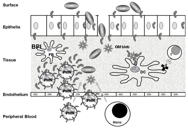
The initial interactions between Gram-negative bacteria, endotoxin and BPI normally takes place in the tissue. BPI is present on the apical and basolateral surfaces of the epithelium but if levels of BPI expression at these sites are great enough to increase local defences against invading GNB is unknown. Fibroblasts located under the epithelial lining also express BPI and could serve as a second barrier against GNB and endotoxin. However, the major source of BPI in tissue is derived from recruited and activated neutrophils. BPI can promote delivery of GNB remnants (e.g. OM blebs) to dendritic cells, facilitating induction of T-cell-dependent antibacterial immunity. BPI expression in fibroblasts and epithelia later in the immune response by IL-4 and lipoxins, respectively [38, 39] may further promote elimination of GNB and resolution of endotoxin-induced inflammation.
Dendritic cells have not only the potential to ingest bacteria but can also internalize and process OM blebs and BPI delivered to them and provide a link to the adaptive immune system. Abbreviations: PMN polymorphonuclear neutrophil granulocyte, GNB Gram-negative bacteria, Mono monocyte, FB fibroblast, DC dendritic cell, BPI bactericidal/permeability increasing protein, OM blebs outer membrane vesicles of GNB.
The activation of host cells by endotoxin requires the extraction and transfer of individual endotoxin molecules to a host cell surface receptor complex containing MD-2 and Toll-like receptor (TLR) 4 [74–76]. This transfer does not occur directly but rather requires first the interaction of LBP with the endotoxin-rich GNB outer membrane and transfer of endotoxin monomers to either soluble or membrane-bound CD14. The monomeric endotoxin:CD14 complex is the preferred substrate for MD-2 – either MD-2 alone or MD-2 pre-associated with TLR4 [74, 75]. Simultaneous binding of endotoxin and TLR4 by MD-2 then triggers potent pro-inflammatory responses (Figure 4A, [74, 75]).
Figure 4. Roles of BPI during GNB infection.

Role of BPI in the interaction of GNB and OM blebs with neutrophil granulocytes and monocytes (A). BPI can opsonize GNB and target them to be phagocytosed by PMN. It can contribute to intracellular killing of ingested GNB and also to extracellular bacterial killing when released to the extracellular space by PMN. Single endotoxin molecules from OM blebs or other endotoxin containing remnants of GNB can be extracted and processed by the LBP/CD14/MD-2/TLR-4 pathway resulting an activation of CD-14 positive monocytes. In contrast, BPI bound endotoxin is not accessible for LBP/CD14 mediated LOS/LPS extraction and does not cause an inflammatory response in monocytes [81, 82, 107].
Role of BPI in the interaction of OM blebs with dendritic cells (B). BPI can promote CD14-independent delivery of OM blebs to dendritic cells (DC). In parallel, LBP and sCD14 can promote MDDC activation by extraction and delivery of endotoxin to MD-2/TLR4 [108]. Subsequently, the ability of BPI and LBP to interact with the same OM bleb could allow coincidental enhancement of antigen delivery by BPI and LBP/sCD14-dependent cell activation and could thereby promote antigen-presenting functions of DC. Abbreviations: BPI Bactericidal/permeability-increasing protein, GNB Gram negative bacteria, LBP Lipopolysaccharides binding protein, CD14 either membrane bound or soluble CD14, Blebs outer membrane vesicles of GNB, LOS/LPS lipooligosaccharide or lipopolysaccharide, MD-2 Myeloid Differentiation-2, TLR-4 TOLL-like receptor-4
LBP is produced constitutively in the liver and thus is constantly present in the circulation and, at lower levels, in tissue. These low levels of LBP are most favorable for extraction and transfer of endotoxin monomers to CD14 and, subsequently, to MD-2/TLR4, especially early after GNB invasion when the bacteria and endotoxin are most likely present in minute amounts. Before induction of acute inflammation, there are no neutrophils within tissue and thus no neutrophil-derived BPI. The extent to which basal expression of BPI in tissue by other cells (e.g., epithelia, connective tissue) could be functionally significant is unclear [38, 39, 42, 77–79]. Tissue-derived BPI has been found on apical and basolateral epithelial surfaces [38, 40, 42] and on dermal fibroblasts [39] derived from subcutaneous connective tissue, however, its quantitative contribution to the initial host interactions with GNB in tissue are as yet unknown. The presence of BPI at these sites could enhance the ability of local defenses to eliminate small amounts of invading bacteria and also diminish the availability of endotoxin for stimulation of sub-epithelial host cells via the LBP/CD14/MD-2/TLR4 pathway [80–82]. Under these circumstances, incidental incursions of GNB may be resolved locally without a significant inflammatory response and little or no influx of neutrophils.
Once, however, a threshold of GNB invasion and endotoxin exposure has been reached, the low levels of LBP in interstitial fluid at rest are likely sufficient to initiate delivery of LPS to mCD14/MD-2/TLR4 on endotoxin responsive resident tissue cells (e.g., macrophages) and potently induce acute inflammation. Effects of acute inflammation include the release of bioactive substances (e.g. cytokines and chemokines) that promote the permeation of antimicrobial plasma factors into the local tissue and recruitment of neutrophils and, later, mononuclear blood cells, monocytes and lymphocytes, to the site of infection. In this setting, recruited neutrophils use much of their granule contents, including BPI, to mediate intracellular cytotoxicity after phagocytosis of the invading bacteria. In addition, recruited and activated neutrophils release some of their granule contents into the surrounding inflammatory fluid [83–86]. The released proteins may contribute to containment and killing of extracellular bacteria that have exceeded or evaded/resisted the phagocytic capacity of the neutrophils and also to elimination of bioactive bacterial remnants [87, 88].
The antibiotic effects of BPI on GNB take place in two stages [89] involving: i) potentially reversible, sub-lethal alterations of the outer membrane; and ii) irreversible lesions extending to the inner cytoplasmic membrane, disrupting energy-generating and -dependent biochemical machinery and resulting in cell death [89, 90]. The sub-lethal effects of BPI can be amplified by synergistic actions with other neutrophil granule proteins [91–95] and with extracellular antimicrobial proteins that are mobilized in neutrophil-rich inflammatory exudates, including group IIA phospholipase A2 (gIIA PLA2) and complement [96–98]. These combined actions can take place both within neutrophils after uptake of the PLA2 and complement with bacteria during phagocytosis [97, 98] and in the extracellular inflammatory fluid [40, 86, 99–101]. Levels of BPI in cell-free inflammatory fluid of neutrophil-rich exudates are functionally significant (≥10 nM) and co-exist with other antimicrobial host factors that can act in concert with BPI [76, 86, 100–104]. Thus, in cooperation with other cellular and humoral components of the innate immune system, BPI can contribute to killing of invading GNB.
During and after phagocytosis, the lipid A region within bacterial endotoxin is partially deacylated by the host enzyme acyloxyacyl hydrolase [88, 105]. Acyloxyacyl hydrolase converts hexa-acylated endotoxins that are potent TLR4 agonists to tetra-acylated species that are TLR4 antagonists [3, 105, 106]. Endotoxin associated with intact bacteria and bacterial remnants is also partially deacylated in the extracellular inflammatory fluid [88] but at a much slower rate. Thus, a more rapid detoxification mechanism may be needed in inflammatory fluids and could be mediated by locally mobilized endotoxin binding proteins, such as BPI [86]. Like LBP, BPI binds to the highly conserved lipid A region of endotoxin [9]. In contrast to the effect of binding of LBP to endotoxin, binding of BPI does not lead to extraction and transfer of endotoxin to CD14 and thus can preclude MD-2/TLR4-dependent cell activation by endotoxin [51, 66]. This difference reflects differences in the structural and functional properties of the C-terminal domain of LBP and of BPI [48, 51, 81, 107]. Additional differences in the structural and functional properties of the N-terminal domain of BPI and of LBP account for the higher affinity of BPI (vs. LBP) for purified or outer membrane-inserted endotoxin [9, 52, 53]. Thus, BPI can effectively compete with LBP for binding to endotoxin and thus inhibit LBP-dependent (and –independent) extraction and transfer of endotoxin to CD14/MD-2/TLR4 [81, 82].
Although BPI binding to aggregates of endotoxin, cell-free outer membrane blebs or intact GNB does not engage CD14-dependent cellular interactions, BPI can promote CD14-independent interactions with specific host cells [48, 51–53]. In each instance, these effects of BPI are dependent on the N-teminal domain of BPI to bind endotoxin and the C-terminal domain of BPI to somehow facilitate targeting of the BPI-coated endotoxin-rich particle to a host cell. Remarkably, which host cell is engaged seems dependent on the nature of the endotoxin-rich particle engaged by BPI. In experiments using encapsulated Neisseria meningitidis serogroup B and cell-free outer membrane blebs and aggregates of endotoxin derived from these bacteria, intact bacteria treated with BPI were delivered to neutrophils > monocytes, purified aggregates of endotoxin treated with BPI were delivered to monocytes > neutrophils and outer membrane blebs to monocytes and monocyte-derived dendritic cells > macrophages [51–53](Schultz et al., manuscript submitted, Figure 4). The molecular bases of the selective association of different BPI-coated endotoxin-rich particles with specific host cells are not known.
Opsonic effects of BPI on phagocytosis by neutrophils of phagocytosis-resistant encapsulated GNB require doses of BPI sufficient to induce sub-lethal injury [52]. Uptake of the BPI-coated bacteria is accompanied by activation of the cellular respiratory burst indicating that the non-oxidative antibacterial action initiated by extracellular BPI will be followed and amplified by oxygen-dependent (and additional oxygen-independent) cytotoxic actions within the neutrophil. In contrast, BPI-dependent uptake of cell-free endotoxin by monocytes is not accompanied by apparent activation of the monocytes. In fact, low levels of endotoxin- and CD14/MD-2/TLR4-dependent activation of the monocytes that are induced by the cell-free endotoxin-rich particles are inhibited by BPI despite increased uptake of endotoxin by the monocytes [51, 53]. These findings are most compatible with a role for BPI in the “silent” disposal of cell-free endotoxin that needs to be removed or detoxified by AOAH to help resolve endotoxin-induced inflammation. However, the magnitude of BPI-dependent cell-free endotoxin clearance that has been observed thus far in in vitro studies has been quite limited (≤ 15% of that present), raising the possibility that additional co-factors, absent in the in vitro assays, may be needed for more efficient BPI-dependent clearance or that disposal is not a major function of BPI-dependent interactions of cell-free endotoxin with host cells.
The recent demonstration (Schultz et al., manuscript submitted) that BPI promotes delivery and uptake of purified endotoxin and, to a greater extent, outer membrane blebs to dendritic cells and not to monocyte-derived macrophages suggests a novel role for BPI in the uptake and presentation of bacterial antigens by professional antigen presenting cells (Figure 4B). Enhanced uptake of the blebs by dendritic cells in the presence of BPI does not increase cell activation but still permits LBP/sCD14-dependent cell activation by the blebs. We speculate that a single bleb containing tens-hundreds of thousands of endotoxin molecules/particle is able to accommodate both BPI- and LBP-dependent interactions, the former increasing uptake of the particles, the latter promoting CD14/MD-2/TLR4-dependent cell activation. According to current concepts [108], initiation of TLR4 signaling from the same intracellular compartment containing internalized bacterial antigens should favor subsequent processing and MHC II-dependent presentation of fragments of the internalized protein antigens. Thus, these findings suggest that innate immune recognition of endotoxin by BPI and LBP can promote, in a complementary manner, induction of anti-bacterial adaptive immunity.
Recent studies have demonstrated that BPI has the ability to induce apoptosis in human vascular endothelial cells in vitro and inhibit their migration in a wound assay [54]. Moreover, the amino-terminal part of BPI (rBPI21) inhibited neo-vascularization in the retina of mice [55, 56], either ischemia-induced or diabetes-induced, suggesting an antiangiogenetic effect of BPI. Its role in the context of a GNB-induced inflammation in a tissue setting remains to be elucidated. It is, however, conceivable that BPI could impair vascular access of GNB by inhibiting neo-angiogenesis in settings where chronic neutrophilic inflammation prevails, e.g. in inflammatory bowel diseases or cystic fibrosis [77, 79, 109, 110].
Autoimmunity against BPI: BPI-ANCA
Anti-neutrophil cytoplasmic autoantibodies (ANCA), represent a heterogenous group of autoantibodies directed against neutrophil granule proteins. The presence of ANCA typically leads to a characteristic staining pattern in human neutrophils, detectable by indirect immunofluorescence microscopy [111]. Originally described in patients with rapid progressive glomerulonephritis, ANCA soon became hallmarks in the diagnostic workup and monitoring of disease activity in primary systemic vasculitides [112]. In Wegener’s granulomatosis and microscopic polyangiitis, there is a strong association with ANCA against proteinase 3 (PR3) and myeloperoxidase (MPO), respectively [111]. BPI was also first described as an ANCA target antigen in vasculitides [113, 114]. However, it soon became apparent that BPI is a target of ANCA in a variety of diseases of different etiologies like cystic fibrosis (CF), inflammatory bowel diseases (IBD), reactive arthritis (ReA), HIV, the recently discovered peptide transporter complex associated with antigen presentation (TAP) deficiency [16–18, 113, 115] and chronic obstructive pulmonary disease (COPD, Schultz, H. and Murphy, T.F., unpublished observation). Thus, BPI-ANCA could not be used as a disease-specific seromarker as PR3-ANCA is for Wegener’s granulomatosis. With the exception of HIV, where the levels and function of CD4+ helper T lymphocytes are greatly disturbed [116], a common feature of BPI-ANCA associated diseases is a chronic and/or profuse exposure of host tissue to GNB and/or their endotoxin and an accompanying neutrophilic inflammation. BPI-autoantibodies have been found in the serum of up to 90% of these patients (Figure 5).
Figure 5. Prevalence of BPI-autoantibodies in selected inflammatory diseases (modified after [125]).

Shown is the prevalence of BPI-ANCA in selected studies of healthy persons and patients with infectious diseases, rheumatic diseases, inflammatory bowel diseases and immunodeficiency [17, 18, 115, 118, 119, 126].
Abbreviations: HIV human immunodeficiency virus; ReA reactive arthritis; TAP transporter associated with antigen presentation
The early appearance of BPI-ANCA in some CF children, even before clinical signs of GNB infection, raised the possibility of a genetic predisposition to BPI-autoimmunity, at least in a subset of individuals with BPI-ANCA [117]. However, the apparent association in most cases of BPI-ANCA with GNB infection and neutrophilic inflammation suggest a greater circumstantial component to this auto-immune manifestation. Epitope mapping data have revealed similarities of BPI epitopes recognized by BPI-ANCA (Table 1) with specific protein sequences of infecting GNB (e.g. outer membrane proteins of Pseudomonas aeruginosa) suggesting that cross-reactivity followed by epitope spreading in the inflammatory setting could be a mechanism for broken tolerance against BPI [118, 119]. The waning of BPI-ANCA in CF patients after lung transplantation and a reduced pulmonary burden of P. aeruginosa [120, 121] is consistent with this hypothesis.
Table 1.
Several BPI auto-antibodies of cystic fibrosis and inflammatory bowel disease patients react with regions on human BPI (shown in upper line, numbers correspond to amino acid residues) that are homologous to limited regions of specific proteins of GNB (shown below corresponding BPI sequence). Data are taken from references [114, 118, 119].
| BPI Sequence Bacterial Sequence | Patients n | Bacterial protein | Origin | % Identity |
|---|---|---|---|---|
| 50 AALQKELKRIKIPD 63 LQKELKR | 5 | Protein (argF- lacZ region) | Escherichia coli | 100 |
| 397 AESDRLVGELRLDR 410 AERDRLIGGLRLDR | 4 | OprC | Pseudomonas aeruginosa | 100 |
| 88 FQLPSSQISMVP 99 LHSSQISM | 4 | IpaD ORF | Shigella dysenteriae | 87 |
| 137 GMSISADLKLGSNPT 151 MSIDADLK | 3 | Invasion Protein | Salmonella enterica | 87 |
| 88 FQLPSSQISMVP 99 LHSSQISM | 3 | Invasion Protein | Shigella flexneri | 87 |
| 99 PNVGLKFSISNAN 111 FAISNAN | 2 | FlgE Protein | Campylobacter jejuni | 85 |
An added possibility is suggested by the BPI-dependent interactions of GNB outer membrane blebs with dendritic cells briefly described above. Although the principal physiological function of these interactions may be increased bacterial antigen delivery to antigen-presenting cells leading to increased induction of adaptive immunity to bacterial outer membrane antigens, the uptake of these blebs is likely to include significant amounts of bound BPI. Thus, under conditions of pronounced and protracted GNB infection, endotoxin elaboration and extracellular BPI mobilization, uptake of BPI-coated blebs (or, perhaps, intact GNB) by dendritic cells may result in sufficient MHC II-dependent presentation of BPI by activated dendritic cells to over-ride the normal tolerance of the human host to BPI, resulting in BPI-ANCA. This mechanism would also be consistent with the diminishing levels of BPI-ANCA in CF patients after lung transplantation and fluctuations in BPI-ANCA levels with changes in clinical and infection status.
The correlation of BPI-ANCA levels in CF and IBD patients indicate that BPI-ANCA might be useful to identify subgroups of patients with higher inflammatory activity and more severe organ damage (Table 2, [16, 117–122]). The ability of BPI-ANCA to interfere with the opsonic [123] and antibiotic [18, 119, 124] actions of BPI has been demonstrated in vitro. In addition, BPI-ANCA can activate TNFα-primed PMNs to produce reactive oxygen species in vitro [117], suggesting an additional pro-inflammatory effect these autoantibodies could have in vivo. The extent to which neutrophil- vs. tissue-derived BPI are targets of BPI-ANCA, distinct BPI auto-antibodies directed at different BPI epitopes and mediate different functional effects and BPI-ANCA contribute to inflammatory disease progression requires further study.
Table 2.
Relation of BPI-ANCA titer to clinical status in cystic fibrosis and inflammatorybowel diseases.
| Cystic Fibrosis | Inflammatory Bowel Diseases |
|---|---|
| ↓ FEV1 and FVC* | ↑ Aggressive course |
| ↑ P. aeruginosa colonization* | ↑ Frequency of diarrhea* |
| ↑ ESR* and CRP* | ↑ Neutrophil count* |
| ↓ Chest X ray score* | ↑ Crohn’s disease activity index (CDAI)* |
| ↓ Chest HRCT score* | ↑ Extraintestinal manifestations* |
| ↓ Albumin* | |
| ↑ ESR* and CRP* |
Shown are significant differences between BPI-ANCA positive and negative patients with cystic fibrosis and inflammatory bowel diseases. Arrows heading upwards indicate a positive correlation, arrows directed downwards indicate an inverse correlation between BPI-ANCA titer and the indicated clinical parameter;
Abbreviations: FEV1 forced expiratory volume in 1 second; FVC functional vital capacity of the lung; ESR Erythrocyte sedimentation rate; CRP C-reactive protein; CDAI Crohn’s disease activity index; HRCT high resolution computed tomography
Outlook
BPI is a pluripotent protein located in neutrophils and tissue that likely plays a pivotal role in host defense against GNB and their endotoxin by means of its antibiotic and endotoxin-neutralizing and –disposing functions. Further studies are needed to understand in more detail how expression of BPI is controlled in non-myeloid tissue cells and how this may help regulate host-GNB interactions in tissue settings. A better understanding of the regulation of BPI expression in epithelial and connective tissue settings may suggest novel approaches to increasing local antibacterial and anti-inflammatory properties that could be especially useful in compromised settings such as burn patients. The ability of BPI to promote the delivery of GNB remnants to dendritic cells while still permitting LBP/CD14-dependent signaling from these same particles suggests an expansion of the roles of BPI from an innate immune effector to an inducer of antibacterial adaptive immunity. This previously unrecognized function of BPI may also create circumstances conducive to elaboration of BPI auto-antibodies. Further insight in the processing of BPI, complexed with GNB and/or OM blebs, by professional antigen presenting cells may also help clarify the genesis of BPI auto-antibodies and, by possible analogy, of other ANCA directed at other antimicrobial granule proteins. These studies may also suggest new ways to enhance host adaptive immune response to important bacterial pathogens (e.g. N. meningitidis serogroup B) and thus facilitate the development of new vaccine approaches.
Acknowledgments
Supported in parts by DFG Grant Schu 1308/1-3, Kompetenznetzwerk Rheuma and Forschungsschwerpunkt ”Körpereigene Infektabwehr”, University of Lübeck, Germany (to H.S.) and by U.S. Public Health Service Grants PO144642 and AI59372 (to J.P.W.).
Footnotes
Publisher's Disclaimer: Disclaimers The authors have no financial conflict of interest.
Publisher's Disclaimer: This is a PDF file of an unedited manuscript that has been accepted for publication. As a service to our customers we are providing this early version of the manuscript. The manuscript will undergo copyediting, typesetting, and review of the resulting proof before it is published in its final citable form. Please note that during the production process errors may be discovered which could affect the content, and all legal disclaimers that apply to the journal pertain.
References
- 1.Alexander C, Rietschel ET. Bacterial lipopolysaccharides and innate immunity. J Endotoxin Res. 2001;7:167–202. [PubMed] [Google Scholar]
- 2.Beutler B, Rietschel ET. Innate immune sensing and its roots: the story of endotoxin. Nat Rev Immunol. 2003;3:169–76. doi: 10.1038/nri1004. [DOI] [PubMed] [Google Scholar]
- 3.Munford RS. Detoxifying endotoxin: time, place and person. J Endotoxin Res. 2005;11:69–84. doi: 10.1179/096805105X35161. [DOI] [PubMed] [Google Scholar]
- 4.Dellinger RP. Inflammation and coagulation: implications for the septic patient. Clin Infect Dis. 2003;36:1259–65. doi: 10.1086/374835. [DOI] [PubMed] [Google Scholar]
- 5.Bainton DF. Neutrophilic leukocyte granules: from structure to function. Adv Exp Med Biol. 1993;336:17–33. doi: 10.1007/978-1-4757-9182-2_3. [DOI] [PubMed] [Google Scholar]
- 6.Dallegri F, Ottonello L. Tissue injury in neutrophilic inflammation. Inflamm Res. 1997;46:382–91. doi: 10.1007/s000110050208. [DOI] [PubMed] [Google Scholar]
- 7.Ganz T. Antimicrobial proteins and peptides in host defense. Semin Respir Infect. 2001;16:4–10. doi: 10.1053/srin.2001.22723. [DOI] [PubMed] [Google Scholar]
- 8.Weiss J, Elsbach P, Olsson I, Odeberg H. Purification and characterization of a potent bactericidal and membrane active protein from the granules of human polymorphonuclear leukocytes. J Biol Chem. 1978;253:2664–72. [PubMed] [Google Scholar]
- 9.Gazzano-Santoro H, Parent JB, Grinna L, Horwitz A, Parsons T, Theofan G, Elsbach P, Weiss J, Conlon PJ. High-affinity binding of the bactericidal/permeability-increasing protein and a recombinant amino-terminal fragment to the lipid A region of lipopolysaccharide. Infect Immun. 1992;60:4754–61. doi: 10.1128/iai.60.11.4754-4761.1992. [DOI] [PMC free article] [PubMed] [Google Scholar]
- 10.Marra MN, Wilde CG, Griffith JE, Snable JL, Scott RW. Bactericidal/permeability-increasing protein has endotoxin-neutralizing activity. J Immunol. 1990;144:662–6. [PubMed] [Google Scholar]
- 11.Lin Y, Ammons WS, Leach WJ, Kung AH. Protective effects of a recombinant N-terminal fragment of bactericidal/permeability increasing protein on endotoxic shock in conscious rabbits. Shock. 1994;2:324–31. doi: 10.1097/00024382-199411000-00005. [DOI] [PubMed] [Google Scholar]
- 12.von der Mohlen MA, Kimmings AN, Wedel NI, Mevissen ML, Jansen J, Friedmann N, Lorenz TJ, Nelson BJ, White ML, Bauer R, et al. Inhibition of endotoxin-induced cytokine release and neutrophil activation in humans by use of recombinant bactericidal/permeability-increasing protein. J Infect Dis. 1995;172:144–51. doi: 10.1093/infdis/172.1.144. [DOI] [PubMed] [Google Scholar]
- 13.Wiezer MJ, Langendoen SI, Meijer C, Bauer RJ, White ML, Carroll SF, Meyer S, Thijs LG, van Leeuwen PA. Pharmacokinetics of a recombinant amino terminal fragment of bactericidal/permeability increasing protein (rBPI21) after liver surgery in rats and humans. Shock. 1998;10:161–6. doi: 10.1097/00024382-199809000-00002. discussion 167–8. [DOI] [PubMed] [Google Scholar]
- 14.Bauer RJ, White ML, Wedel N, Nelson BJ, Friedmann N, Cohen A, Hustinx WN, Kung AH. A phase I safety and pharmacokinetic study of a recombinant amino terminal fragment of bactericidal/permeability-increasing protein in healthy male volunteers. Shock. 1996;5:91–6. doi: 10.1097/00024382-199602000-00002. [DOI] [PubMed] [Google Scholar]
- 15.Demetriades D, Smith JS, Jacobson LE, Moncure M, Minei J, Nelson BJ, Scannon PJ. Bactericidal/permeability-increasing protein (rBPI21) in patients with hemorrhage due to trauma: results of a multicenter phase II clinical trial. rBPI21 Acute Hemorrhagic Trauma Study Group. J Trauma. 1999;46:667–76. doi: 10.1097/00005373-199904000-00018. discussion 676–7. [DOI] [PubMed] [Google Scholar]
- 16.Zhao MH, Jayne DR, Ardiles LG, Culley F, Hodson ME, Lockwood CM. Autoantibodies against bactericidal/permeability-increasing protein in patients with cystic fibrosis. Qjm. 1996;89:259–65. doi: 10.1093/qjmed/89.4.259. [DOI] [PubMed] [Google Scholar]
- 17.Stoffel MP, Csernok E, Herzberg C, Johnson T, Carroll SF, Gross WL. Anti-neutrophil cytoplasmic antibodies (ANCA) directed against bactericidal/permeability increasing protein (BPI): a new seromarker for inflammatory bowel disease and associated disorders. Clin Exp Immunol. 1996;104:54–9. doi: 10.1046/j.1365-2249.1996.d01-654.x. [DOI] [PMC free article] [PubMed] [Google Scholar]
- 18.Schultz H, Schinke S, Weiss J, Cerundolo V, Gross WL, Gadola S. BPI-ANCA in transporter associated with antigen presentation (TAP) deficiency: possible role in susceptibility to Gram-negative bacterial infections. Clin Exp Immunol. 2003;133:252–9. doi: 10.1046/j.1365-2249.2003.02197.x. [DOI] [PMC free article] [PubMed] [Google Scholar]
- 19.Kirschning CJ, Au-Young J, Lamping N, Reuter D, Pfeil D, Seilhamer JJ, Schumann RR. Similar organization of the lipopolysaccharide-binding protein (LBP) and phospholipid transfer protein (PLTP) genes suggests a common gene family of lipid-binding proteins. Genomics. 1997;46:416–25. doi: 10.1006/geno.1997.5030. [DOI] [PubMed] [Google Scholar]
- 20.Schumann RR, Leong SR, Flaggs GW, Gray PW, Wright SD, Mathison JC, Tobias PS, Ulevitch RJ. Structure and function of lipopolysaccharide binding protein. Science. 1990;249:1429–31. doi: 10.1126/science.2402637. [DOI] [PubMed] [Google Scholar]
- 21.Bingle CD, Craven CJ. PLUNC: a novel family of candidate host defence proteins expressed in the upper airways and nasopharynx. Hum Mol Genet. 2002;11:937–43. doi: 10.1093/hmg/11.8.937. [DOI] [PubMed] [Google Scholar]
- 22.Hubacek JA, Buchler C, Aslanidis C, Schmitz G. The genomic organization of the genes for human lipopolysaccharide binding protein (LBP) and bactericidal permeability increasing protein (BPI) is highly conserved. Biochem Biophys Res Commun. 1997;236:427–30. doi: 10.1006/bbrc.1997.6970. [DOI] [PubMed] [Google Scholar]
- 23.Larsen K, Madsen LB, Bendixen C, Klein W, Tromm A, Folwaczny C, Hagedorn M, Duerig N, Epplen J, Schmiegel W, Griga T. Porcine SPLUNC1: Molecular cloning, characterization and expression analysis A Polymorphism of the Bactericidal/Permeability Increasing Protein (BPI) Gene Is Associated With Crohn’s Disease. Biochim Biophys Acta. 2005;39:282–283. [Google Scholar]
- 24.Gullberg U, Andersson E, Garwicz D, Lindmark A, Olsson I. Biosynthesis, processing and sorting of neutrophil proteins: insight into neutrophil granule development. Eur J Haematol. 1997;58:137–53. doi: 10.1111/j.1600-0609.1997.tb00940.x. [DOI] [PubMed] [Google Scholar]
- 25.Lennartsson A, Pieters K, Vidovic K, Gullberg U. A murine antibacterial ortholog to human bactericidal/permeability-increasing protein (BPI) is expressed in testis, epididymis, and bone marrow. J Leukoc Biol. 2005;77:369–77. doi: 10.1189/jlb.0304159. [DOI] [PubMed] [Google Scholar]
- 26.Eckert M, Wittmann I, Rollinghoff M, Gessner A, Schnare M. Endotoxin-induced expression of murine bactericidal permeability/increasing protein is mediated exclusively by toll/IL-1 receptor domain-containing adaptor inducing IFN-beta-dependent pathways. J Immunol. 2006;176:522–8. doi: 10.4049/jimmunol.176.1.522. [DOI] [PubMed] [Google Scholar]
- 27.Kono T, Sakai M. Molecular cloning of a novel bactericidal permeability-increasing protein/lipopolysaccharide-binding protein (BPI/LBP) from common carp Cyprinus carpio L. and its expression. Mol Immunol. 2003;40:269–78. doi: 10.1016/s0161-5890(03)00103-2. [DOI] [PubMed] [Google Scholar]
- 28.Inagawa H, Honda T, Kohchi C, Nishizawa T, Yoshiura Y, Nakanishi T, Yokomizo Y, Soma G. Cloning and characterization of the homolog of mammalian lipopolysaccharide-binding protein and bactericidal permeability-increasing protein in rainbow trout Oncorhynchus mykiss. J Immunol. 2002;168:5638–44. doi: 10.4049/jimmunol.168.11.5638. [DOI] [PubMed] [Google Scholar]
- 29.Stenvik J, Solstad T, Strand C, Leiros I, Jorgensen TT. Cloning and analyses of a BPI/LBP cDNA of the Atlantic cod (Gadus morhua L.) Dev Comp Immunol. 2004;28:307–23. doi: 10.1016/j.dci.2003.09.002. [DOI] [PubMed] [Google Scholar]
- 30.Calafat J, Janssen H, Tool A, Dentener MA, Knol EF, Rosenberg HF, Egesten A. The bactericidal/permeability-increasing protein (BPI) is present in specific granules of human eosinophils. Blood. 1998;91:4770–5. [PubMed] [Google Scholar]
- 31.Dentener MA, Francot GJ, Buurman WA. Bactericidal/permeability-increasing protein, a lipopolysaccharide-specific protein on the surface of human peripheral blood monocytes. J Infect Dis. 1996;173:252–5. doi: 10.1093/infdis/173.1.252. [DOI] [PubMed] [Google Scholar]
- 32.Weersink AJ, van Kessel KP, van den Tol ME, van Strijp JA, Torensma R, Verhoef J, Elsbach P, Weiss J. Human granulocytes express a 55-kDa lipopolysaccharide-binding protein on the cell surface that is identical to the bactericidal/permeability-increasing protein. J Immunol. 1993;150:253–63. [PubMed] [Google Scholar]
- 33.Weiss J, Olsson I. Cellular and subcellular localization of the bactericidal/permeability-increasing protein of neutrophils. Blood. 1987;69:652–9. [PubMed] [Google Scholar]
- 34.Calafat J, Janssen H, Knol EF, Malm J, Egesten A. The bactericidal/permeability-increasing protein (BPI) is membrane-associated in azurophil granules of human neutrophils, and relocation occurs upon cellular activation. Apmis. 2000;108:201–8. doi: 10.1034/j.1600-0463.2000.d01-45.x. [DOI] [PubMed] [Google Scholar]
- 35.Lennartsson A, Pieters K, Ullmark T, Vidovic K, Gullberg U. AML-1, PU.1, and Sp3 regulate expression of human bactericidal/permeability-increasing protein. Biochem Biophys Res Commun. 2003;311:853–63. doi: 10.1016/j.bbrc.2003.10.067. [DOI] [PubMed] [Google Scholar]
- 36.Lennartsson A, Vidovic K, Pass MB, Cowland JB, Gullberg U. All-trans retinoic acid-induced expression of bactericidal/permeability-increasing protein (BPI) in human myeloid cells correlates to binding of C/EBPbeta and C/EBPepsilon to the BPI promoter. J Leukoc Biol. 2006;80:196–203. doi: 10.1189/jlb.1205759. [DOI] [PubMed] [Google Scholar]
- 37.Levy O, Martin S, Eichenwald E, Ganz T, Valore E, Carroll SF, Lee K, Goldmann D, Thorne GM. Impaired innate immunity in the newborn: newborn neutrophils are deficient in bactericidal/permeability-increasing protein. Pediatrics. 1999;104:1327–33. doi: 10.1542/peds.104.6.1327. [DOI] [PubMed] [Google Scholar]
- 38.Canny G, Levy O, Furuta GT, Narravula-Alipati S, Sisson RB, Serhan CN, Colgan SP. Lipid mediator-induced expression of bactericidal/permeability-increasing protein (BPI) in human mucosal epithelia. Proc Natl Acad Sci U S A. 2002;99:3902–7. doi: 10.1073/pnas.052533799. [DOI] [PMC free article] [PubMed] [Google Scholar]
- 39.Reichel PH, Seemann C, Csernok E, Schroder JM, Muller A, Gross WL, Schultz H. Bactericidal/permeability-increasing protein is expressed by human dermal fibroblasts and upregulated by interleukin 4. Clin Diagn Lab Immunol. 2003;10:473–5. doi: 10.1128/CDLI.10.3.473-475.2003. [DOI] [PMC free article] [PubMed] [Google Scholar]
- 40.Paulsen FP, Pufe T, Schaudig U, Held-Feindt J, Lehmann J, Schroder JM, Tillmann BN. Detection of natural peptide antibiotics in human nasolacrimal ducts. Invest Ophthalmol Vis Sci. 2001;42:2157–63. [PubMed] [Google Scholar]
- 41.Peuravuori H, Aho VV, Aho HJ, Collan Y, Saari KM. Bactericidal/permeability-increasing protein in lacrimal gland and in tears of healthy subjects. Graefes Arch Clin Exp Ophthalmol. 2006;244:143–8. doi: 10.1007/s00417-005-0062-z. [DOI] [PubMed] [Google Scholar]
- 42.Canny GO, Trifonova RT, Kindelberger DW, Colgan SP, Fichorova RN. Expression and function of bactericidal/permeability-increasing protein in human genital tract epithelial cells. J Infect Dis. 2006;194:498–502. doi: 10.1086/505712. [DOI] [PubMed] [Google Scholar]
- 43.Serhan CN. Resolution Phases of Inflammation: Novel Endogenous Anti-Inflammatory and Proresolving Lipid Mediators and Pathways. Annu Rev Immunol. 2006 doi: 10.1146/annurev.immunol.25.022106.141647. [DOI] [PubMed] [Google Scholar]
- 44.Serhan CN, Chiang N. Lipid-derived mediators in endogenous anti-inflammation and resolution: lipoxins and aspirin-triggered 15-epi-lipoxins. Scientific World Journal. 2002;2:169–204. doi: 10.1100/tsw.2002.81. [DOI] [PMC free article] [PubMed] [Google Scholar]
- 45.Ooi CE, Weiss J, Elsbach P, Frangione B, Mannion B. A 25-kDa NH2-terminal fragment carries all the antibacterial activities of the human neutrophil 60-kDa bactericidal/permeability-increasing protein. J Biol Chem. 1987;262:14891–4. [PubMed] [Google Scholar]
- 46.Beamer LJ, Carroll SF, Eisenberg D. Crystal structure of human BPI and two bound phospholipids at 2.4 angstrom resolution. Science. 1997;276:1861–4. doi: 10.1126/science.276.5320.1861. [DOI] [PubMed] [Google Scholar]
- 47.Gazzano-Santoro H, Parent JB, Conlon PJ, Kasler HG, Tsai CM, Lill-Elghanian DA, Hollingsworth RI. Characterization of the structural elements in lipid A required for binding of a recombinant fragment of bactericidal/permeability-increasing protein rBPI23. Infect Immun. 1995;63:2201–5. doi: 10.1128/iai.63.6.2201-2205.1995. [DOI] [PMC free article] [PubMed] [Google Scholar]
- 48.Abrahamson SL, Wu HM, Williams RE, Der K, Ottah N, Little R, Gazzano-Santoro H, Theofan G, Bauer R, Leigh S, Orme A, Horwitz AH, Carroll SF, Dedrick RL. Biochemical characterization of recombinant fusions of lipopolysaccharide binding protein and bactericidal/permeability-increasing protein. Implications in biological activity. J Biol Chem. 1997;272:2149–55. doi: 10.1074/jbc.272.4.2149. [DOI] [PubMed] [Google Scholar]
- 49.Ooi CE, Weiss J, Doerfler ME, Elsbach P. Endotoxin-neutralizing properties of the 25 kD N-terminal fragment and a newly isolated 30 kD C-terminal fragment of the 55–60 kD bactericidal/permeability-increasing protein of human neutrophils. J Exp Med. 1991;174:649–55. doi: 10.1084/jem.174.3.649. [DOI] [PMC free article] [PubMed] [Google Scholar]
- 50.Horwitz AH, Ammons WS, Bauer RJ, Dedrick R, Nadell R, Williams RE, Liu PS. rBPI(10–193) is secreted by CHO cells and retains the activity of rBPI21. J Endotoxin Res. 2004;10:97–106. doi: 10.1179/096805104225003843. [DOI] [PubMed] [Google Scholar]
- 51.Iovine N, Eastvold J, Elsbach P, Weiss JP, Gioannini TL. The carboxyl-terminal domain of closely related endotoxin-binding proteins determines the target of protein-lipopolysaccharide complexes. J Biol Chem. 2002;277:7970–8. doi: 10.1074/jbc.M109622200. [DOI] [PubMed] [Google Scholar]
- 52.Iovine NM, Elsbach P, Weiss J. An opsonic function of the neutrophil bactericidal/permeability-increasing protein depends on both its N- and C-terminal domains. Proc Natl Acad Sci U S A. 1997;94:10973–8. doi: 10.1073/pnas.94.20.10973. [DOI] [PMC free article] [PubMed] [Google Scholar]
- 53.Hume JR. Department of Microbiology. The University of Iowa; Iowa City: 2005. BPI-mediated delivery of endotoxin-rich particles to human CD14+ monocytes: molecular determinants and fate of endotoxin; p. 165. [Google Scholar]
- 54.van der Schaft DW, Toebes EA, Haseman JR, Mayo KH, Griffioen AW. Bactericidal/permeability-increasing protein (BPI) inhibits angiogenesis via induction of apoptosis in vascular endothelial cells. Blood. 2000;96:176–81. [PubMed] [Google Scholar]
- 55.Yamagata M, Rook SL, Sassa Y, Ma RC, Geraldes P, Goddard L, Clermont A, Gao B, Salti H, Gundel R, White M, Feener EP, Aiello LP, King GL. Bactericidal/permeability-increasing protein’s signaling pathways and its retinal trophic and anti-angiogenic effects. Faseb J. 2006;20:2058–67. doi: 10.1096/05-5662com. [DOI] [PubMed] [Google Scholar]
- 56.Rauniyar RK, Suzuma K, King AL, Aiello LP, King GL. Differential effects of bactericidal/permeability-increasing protein (BPI) analogues on retinal neovascularization and retinal pericyte growth. Invest Ophthalmol Vis Sci. 2002;43:503–9. [PubMed] [Google Scholar]
- 57.Nishimura H, Gogami A, Miyagawa Y, Nanbo A, Murakami Y, Baba T, Nagasawa S. Bactericidal/permeability-increasing protein promotes complement activation for neutrophil-mediated phagocytosis on bacterial surface. Immunology. 2001;103:519–25. doi: 10.1046/j.1365-2567.2001.01263.x. [DOI] [PMC free article] [PubMed] [Google Scholar]
- 58.Lechner AJ, Lamprech KE, Johanns CA, Matuschak GM. The recombinant 23-kDa N-terminal fragment of bactericidal/permeability-increasing protein (rBPI23) decreases Escherichia coli-induced mortality and organ injury during immunosuppression-related neutropenia. Shock. 1995;4:298–306. doi: 10.1097/00024382-199510000-00012. [DOI] [PubMed] [Google Scholar]
- 59.Evans TJ, Carpenter A, Moyes D, Martin R, Cohen J. Protective effects of a recombinant amino-terminal fragment of human bactericidal/permeability-increasing protein in an animal model of gram-negative sepsis. J Infect Dis. 1995;171:153–60. doi: 10.1093/infdis/171.1.153. [DOI] [PubMed] [Google Scholar]
- 60.Alexander S, Bramson J, Foley R, Xing Z. Protection from endotoxemia by adenoviral-mediated gene transfer of human bactericidal/permeability-increasing protein. Blood. 2004;103:93–9. doi: 10.1182/blood-2003-02-0660. [DOI] [PubMed] [Google Scholar]
- 61.Jellema WT, Veerman DP, De Winter RJ, Wesseling KH, Van Deventer SJ, Hack CE, van Lieshout JJ. In vivo interaction of endotoxin and recombinant bactericidal/permeability-increasing protein (rBPI23): hemodynamic effects in a human endotoxemia model. J Lab Clin Med. 2002;140:228–35. doi: 10.1067/mlc.2002.127170. [DOI] [PubMed] [Google Scholar]
- 62.Thomas JA, Tsen MF, White DJ, Horton JW. TLR4 inactivation and rBPI(21) block burn-induced myocardial contractile dysfunction. Am J Physiol Heart Circ Physiol. 2002;283:H1645–55. doi: 10.1152/ajpheart.01107.2001. [DOI] [PubMed] [Google Scholar]
- 63.Levin M, Quint PA, Goldstein B, Barton P, Bradley JS, Shemie SD, Yeh T, Kim SS, Cafaro DP, Scannon PJ, Giroir BP. Recombinant bactericidal/permeability-increasing protein (rBPI21) as adjunctive treatment for children with severe meningococcal sepsis: a randomised trial. rBPI21 Meningococcal Sepsis Study Group. Lancet. 2000;356:961–7. doi: 10.1016/s0140-6736(00)02712-4. [DOI] [PubMed] [Google Scholar]
- 64.Giroir BP, Quint PA, Barton P, Kirsch EA, Kitchen L, Goldstein B, Nelson BJ, Wedel NJ, Carroll SF, Scannon PJ. Preliminary evaluation of recombinant amino-terminal fragment of human bactericidal/permeability-increasing protein in children with severe meningococcal sepsis. Lancet. 1997;350:1439–43. doi: 10.1016/s0140-6736(97)06468-4. [DOI] [PubMed] [Google Scholar]
- 65.Nathan C. Points of control in inflammation. Nature. 2002;420:846–52. doi: 10.1038/nature01320. [DOI] [PubMed] [Google Scholar]
- 66.Weiss JJ. Bactericidal/permeability-increasing protein (BPI) and lipopolysaccharide-binding protein (LBP): structure, function and regulation in host defence against Gram-negative bacteria. Transactions. 2003;31:785–790. doi: 10.1042/bst0310785. [DOI] [PubMed] [Google Scholar]
- 67.Lu M, Zhang M, Takashima A, Weiss J, Apicella MA, Li XH, Yuan D, Munford RS. Lipopolysaccharide deacylation by an endogenous lipase controls innate antibody responses to Gram-negative bacteria. Nat Immunol. 2005;6:989–94. doi: 10.1038/ni1246. [DOI] [PubMed] [Google Scholar]
- 68.Devoe IW, Gilchrist JE. Release of endotoxin in the form of cell wall blebs during in vitro growth of Neisseria meningitidis. J Exp Med. 1973;138:1156–67. doi: 10.1084/jem.138.5.1156. [DOI] [PMC free article] [PubMed] [Google Scholar]
- 69.Post DM, Zhang D, Eastvold JS, Teghanemt A, Gibson BW, Weiss JP. Biochemical and functional characterization of membrane blebs purified from Neisseria meningitidis serogroup B. J Biol Chem. 2005;280:38383–94. doi: 10.1074/jbc.M508063200. [DOI] [PubMed] [Google Scholar]
- 70.Munford RS, Hall CL, Lipton JM, Dietschy JM. Biological activity, lipoprotein-binding behavior, and in vivo disposition of extracted and native forms of Salmonella typhimurium lipopolysaccharides. J Clin Invest. 1982;70:877–88. doi: 10.1172/JCI110684. [DOI] [PMC free article] [PubMed] [Google Scholar]
- 71.Al-Bader T, Jolley KA, Humphries HE, Holloway J, Heckels JE, Semper AE, Friedmann PS, Christodoulides M. Activation of human dendritic cells by the PorA protein of Neisseria meningitidis. Cell Microbiol. 2004;6:651–62. doi: 10.1111/j.1462-5822.2004.00392.x. [DOI] [PubMed] [Google Scholar]
- 72.Sierra GV, Campa HC, Varcacel NM, Garcia IL, Izquierdo PL, Sotolongo PF, Casanueva GV, Rico CO, Rodriguez CR, Terry MH. Vaccine against group B Neisseria meningitidis: protection trial and mass vaccination results in Cuba. NIPH Ann. 1991;14:195–207. discussion 208–10. [PubMed] [Google Scholar]
- 73.Wong S, Lennon D, Jackson C, Stewart J, Reid S, Crengle S, Tilman S, Aaberge I, O’Hallahan J, Oster P, Mulholland K, Martin D. New Zealand Epidemic Strain Meningococcal B Outer Membrane Vesicle Vaccine in Children Aged 16–24 Months. Pediatr Infect Dis J. 2007;26:345–350. doi: 10.1097/01.inf.0000258697.05341.2c. [DOI] [PubMed] [Google Scholar]
- 74.Gioannini TL, Teghanemt A, Zhang D, Levis EN, Weiss JP. Monomeric endotoxin:protein complexes are essential for TLR4-dependent cell activation. J Endotoxin Res. 2005;11:117–23. doi: 10.1179/096805105X35198. [DOI] [PubMed] [Google Scholar]
- 75.Prohinar P, Re F, Widstrom R, Zhang D, Teghanemt A, Weiss JP, Gioannini TL. Specific High Affinity Interactions of Monomeric Endotoxin: Protein Complexes with Toll-like Receptor 4 Ectodomain. J Biol Chem. 2007;282:1010–7. doi: 10.1074/jbc.M609400200. [DOI] [PubMed] [Google Scholar]
- 76.Opal SM, Palardy JE, Marra MN, Fisher CJ, Jr, McKelligon BM, Scott RW. Relative concentrations of endotoxin-binding proteins in body fluids during infection. Lancet. 1994;344:429–31. doi: 10.1016/s0140-6736(94)91767-1. [DOI] [PubMed] [Google Scholar]
- 77.Monajemi H, Meenan J, Lamping R, Obradov D, Radema S, Trown P, Tytgat G, Van Deventer S. Inflammatory bowel disease is associated with increased mucosal levels of bactericidal/permeability-increasing protein. Gastroenterology. 1996;110:733–739. doi: 10.1053/gast.1996.v110.pm8608882. [DOI] [PubMed] [Google Scholar]
- 78.Paulsen F, Pufe T, Conradi L, Varoga D, Tsokos M, Papendieck J, Petersen W. Antimicrobial peptides are expressed and produced in healthy and inflamed human synovial membranes. J Pathol. 2002;198:369–77. doi: 10.1002/path.1224. [DOI] [PubMed] [Google Scholar]
- 79.Haapamaki MM, Haggblom JO, Gronroos JM, Pekkala E, Alanen K, Nevalainen TJ. Bactericidal/permeability-increasing protein in colonic mucosa in ulcerative colitis. Hepatogastroenterology. 1999;46:2273–7. [PubMed] [Google Scholar]
- 80.Dentener MA, Von Asmuth EJ, Francot GJ, Marra MN, Buurman WA. Antagonistic effects of lipopolysaccharide binding protein and bactericidal/permeability-increasing protein on lipopolysaccharide-induced cytokine release by mononuclear phagocytes. Competition for binding to lipopolysaccharide. J Immunol. 1993;151:4258–65. [PubMed] [Google Scholar]
- 81.Heumann D, Gallay P, Betz-Corradin S, Barras C, Baumgartner JD, Glauser MP. Competition between bactericidal/permeability-increasing protein and lipopolysaccharide-binding protein for lipopolysaccharide binding to monocytes. J Infect Dis. 1993;167:1351–7. doi: 10.1093/infdis/167.6.1351. [DOI] [PubMed] [Google Scholar]
- 82.Gazzano-Santoro H, Meszaros K, Birr C, Carroll SF, Theofan G, Horwitz AH, Lim E, Aberle S, Kasler H, Parent JB. Competition between rBPI23, a recombinant fragment of bactericidal/permeability-increasing protein, and lipopolysaccharide (LPS)-binding protein for binding to LPS and gram-negative bacteria. Infect Immun. 1994;62:1185–91. doi: 10.1128/iai.62.4.1185-1191.1994. [DOI] [PMC free article] [PubMed] [Google Scholar]
- 83.Dallegri F, F|Ottonello LL. Tissue injury in neutrophilic inflammation. Inflammation research. 1997;46:382–391. doi: 10.1007/s000110050208. [DOI] [PubMed] [Google Scholar]
- 84.Svanborg C, Godaly G, Hedlund M. Cytokine responses during mucosal infections: role in disease pathogenesis and host defence. Curr Opin Microbiol. 1999;2:99–105. doi: 10.1016/s1369-5274(99)80017-4. [DOI] [PubMed] [Google Scholar]
- 85.Wilson ME. Effects of bacterial endotoxins on neutrophil function. Rev Infect Dis. 1985;7:404–18. doi: 10.1093/clinids/7.3.404. [DOI] [PubMed] [Google Scholar]
- 86.Weinrauch Y, Foreman A, Shu C, Zarember K, Levy O, Elsbach P, Weiss J. Extracellular accumulation of potently microbicidal bactericidal/permeability-increasing protein and p15s in an evolving sterile rabbit peritoneal inflammatory exudate. J Clin Invest. 1995;95:1916–24. doi: 10.1172/JCI117873. [DOI] [PMC free article] [PubMed] [Google Scholar]
- 87.Weinrauch Y, Katz SS, Munford RS, Elsbach P, Weiss J. Deacylation of purified lipopolysaccharides by cellular and extracellular components of a sterile rabbit peritoneal inflammatory exudate. Infect Immun. 1999;67:3376–82. doi: 10.1128/iai.67.7.3376-3382.1999. [DOI] [PMC free article] [PubMed] [Google Scholar]
- 88.Katz SS, Weinrauch Y, Munford RS, Elsbach P, Weiss J. Deacylation of lipopolysaccharide in whole Escherichia coli during destruction by cellular and extracellular components of a rabbit peritoneal inflammatory exudate. J Biol Chem. 1999;274:36579–84. doi: 10.1074/jbc.274.51.36579. [DOI] [PubMed] [Google Scholar]
- 89.Mannion BA, Weiss J, Elsbach P. Separation of sublethal and lethal effects of the bactericidal/permeability increasing protein on Escherichia coli. J Clin Invest. 1990;85:853–60. doi: 10.1172/JCI114512. [DOI] [PMC free article] [PubMed] [Google Scholar]
- 90.in’t Veld G, Mannion B, Weiss J, Elsbach P. Effects of the bactericidal/permeability-increasing protein of polymorphonuclear leukocytes on isolated bacterial cytoplasmic membrane vesicles. Infect Immun. 1988;56:1203–8. doi: 10.1128/iai.56.5.1203-1208.1988. [DOI] [PMC free article] [PubMed] [Google Scholar]
- 91.Zarember K, Elsbach P, Shin-Kim K, Weiss J. p15s (15-kD antimicrobial proteins) are stored in the secondary granules of Rabbit granulocytes: implications for antibacterial synergy with the bactericidal/permeability-increasing protein in inflammatory fluids. Blood. 1997;89:672–9. [PubMed] [Google Scholar]
- 92.Levy O, Weiss J, Ooi CE, Elsbach P. Structural characterization of BPI-modulating 15 kDa proteins from rabbit polymorphonuclear leukocytes: identification of a novel family of leukocyte proteins. Agents Actions. 1993;39(Spec No):C207–10. doi: 10.1007/BF01972768. [DOI] [PubMed] [Google Scholar]
- 93.Levy O, Ooi CE, Weiss J, Lehrer RI, Elsbach P. Individual and synergistic effects of rabbit granulocyte proteins on Escherichia coli. J Clin Invest. 1994;94:672–82. doi: 10.1172/JCI117384. [DOI] [PMC free article] [PubMed] [Google Scholar]
- 94.Levy O, Ooi CE, Elsbach P, Doerfler ME, Lehrer RI, Weiss J. Antibacterial proteins of granulocytes differ in interaction with endotoxin. Comparison of bactericidal/permeability-increasing protein, p15s, and defensins. J Immunol. 1995;154:5403–10. [PubMed] [Google Scholar]
- 95.Zhao H, Kinnunen PK. Modulation of the activity of secretory phospholipase A2 by antimicrobial peptides. Antimicrob Agents Chemother. 2003;47:965–71. doi: 10.1128/AAC.47.3.965-971.2003. [DOI] [PMC free article] [PubMed] [Google Scholar]
- 96.Forst S, Weiss J, Maraganore JM, Heinrikson RL, Elsbach P. Relation between binding and the action of phospholipases A2 on Escherichia coli exposed to the bactericidal/permeability-increasing protein of neutrophils. Biochim Biophys Acta. 1987;920:221–5. doi: 10.1016/0005-2760(87)90098-1. [DOI] [PubMed] [Google Scholar]
- 97.Madsen LM, Inada M, Weiss J. Determinants of activation by complement of group II phospholipase A2 acting against Escherichia coli. Infect Immun. 1996;64:2425–30. doi: 10.1128/iai.64.7.2425-2430.1996. [DOI] [PMC free article] [PubMed] [Google Scholar]
- 98.Inada M, Crowl RM, Bekkers AC, Verheij H, Weiss J. Determinants of the inhibitory action of purified 14-kDa phospholipases A2 on cell-free prothrombinase complex. J Biol Chem. 1994;269:26338–43. [PubMed] [Google Scholar]
- 99.Forst S, Weiss J, Elsbach P, Maraganore JM, Reardon I, Heinrikson RL. Structural and functional properties of a phospholipase A2 purified from an inflammatory exudate. Biochemistry. 1986;25:8381–5. doi: 10.1021/bi00374a008. [DOI] [PubMed] [Google Scholar]
- 100.Aho HJ, Grenman R, Sipila J, Peuravuori H, Hartikainen J, Nevalainen TJ. Group II phospholipase A2 in nasal fluid, mucosa and paranasal sinuses. Acta Otolaryngol. 1997;117:860–3. doi: 10.3109/00016489709114215. [DOI] [PubMed] [Google Scholar]
- 101.Aho VV, Holopainen JM, Tervo T, Moilanen JA, Nevalainen T, Saari KM. Group IIA phospholipase A(2) content in tears of patients having photorefractive keratectomy. J Cataract Refract Surg. 2003;29:2163–7. doi: 10.1016/s0886-3350(03)00419-x. [DOI] [PubMed] [Google Scholar]
- 102.Espinoza J, Chaiworapongsa T, Romero R, Edwin S, Rathnasabapathy C, Gomez R, Bujold E, Camacho N, Kim YM, Hassan S, Blackwell S, Whitty J, Berman S, Redman M, Yoon BH, Sorokin Y. Antimicrobial peptides in amniotic fluid: defensins, calprotectin and bacterial/permeability-increasing protein in patients with microbial invasion of the amniotic cavity, intra-amniotic inflammation, preterm labor and premature rupture of membranes. J Matern Fetal Neonatal Med. 2003;13:2–21. doi: 10.1080/jmf.13.1.2.21. [DOI] [PubMed] [Google Scholar]
- 103.Femling JK, Nauseef WM, Weiss JP. Synergy between extracellular group IIA phospholipase A2 and phagocyte NADPH oxidase in digestion of phospholipids of Staphylococcus aureus ingested by human neutrophils. J Immunol. 2005;175:4653–61. doi: 10.4049/jimmunol.175.7.4653. [DOI] [PubMed] [Google Scholar]
- 104.Inman RD, Chiu B. Comparative microbicidal activity of synovial fluid on arthritogenic organisms. Clin Exp Immunol. 1996;104:80–5. doi: 10.1046/j.1365-2249.1996.d01-650.x. [DOI] [PMC free article] [PubMed] [Google Scholar]
- 105.Munford RS, Hall CL. Detoxification of bacterial lipopolysaccharides (endotoxins) by a human neutrophil enzyme. Science. 1986;234:203–5. doi: 10.1126/science.3529396. [DOI] [PubMed] [Google Scholar]
- 106.Gioannini TL, Teghanemt A, Zhang D, Prohinar P, Levis EN, Munford RS, Weiss JP. Endotoxin binding proteins modulate the susceptibility of bacterial endotoxin to deacylation by acyloxyacyl hydrolase. J Biol Chem. 2007 doi: 10.1074/jbc.M605031200. [DOI] [PubMed] [Google Scholar]
- 107.Tobias PS, Soldau K, Iovine NM, Elsbach P, Weiss J. Lipopolysaccharide (LPS)-binding proteins BPI and LBP form different types of complexes with LPS. J Biol Chem. 1997;272:18682–5. doi: 10.1074/jbc.272.30.18682. [DOI] [PubMed] [Google Scholar]
- 108.Blander JM, Medzhitov R. Toll-dependent selection of microbial antigens for presentation by dendritic cells. Nature. 2006;440:808–12. doi: 10.1038/nature04596. [DOI] [PubMed] [Google Scholar]
- 109.Rastogi D, Ratner AJ, Prince A. Host-bacterial interactions in the initiation of inflammation. Paediatr Respir Rev. 2001;2:245–52. doi: 10.1053/prrv.2001.0147. [DOI] [PubMed] [Google Scholar]
- 110.Aichele D, Schnare M, Saake M, Rollinghoff M, Gessner A. Expression and antimicrobial function of bactericidal permeability-increasing protein in cystic fibrosis patients. Infect Immun. 2006;74:4708–14. doi: 10.1128/IAI.02066-05. [DOI] [PMC free article] [PubMed] [Google Scholar]
- 111.Hoffman GS, Specks U. Antineutrophil cytoplasmic antibodies. Arthritis Rheum. 1998;41:1521–37. doi: 10.1002/1529-0131(199809)41:9<1521::AID-ART2>3.0.CO;2-A. [DOI] [PubMed] [Google Scholar]
- 112.van der Woude FJ, Rasmussen N, Lobatto S, Wiik A, Permin H, van Es LA, van der Giessen M, van der Hem GK, The TH. Autoantibodies against neutrophils and monocytes: tool for diagnosis and marker of disease activity in Wegener’s granulomatosis. Lancet. 1985;1:425–9. doi: 10.1016/s0140-6736(85)91147-x. [DOI] [PubMed] [Google Scholar]
- 113.Zhao MH, Jones SJ, Lockwood CM. Bactericidal/permeability-increasing protein (BPI) is an important antigen for anti-neutrophil cytoplasmic autoantibodies (ANCA) in vasculitis. Clin Exp Immunol. 1995;99:49–56. doi: 10.1111/j.1365-2249.1995.tb03471.x. [DOI] [PMC free article] [PubMed] [Google Scholar]
- 114.Schultz H, Heintz H, van Zandbergen G, Ullrich S, Reinhold-Keller E, Gross WL. ANCA against the bactericidal/permeability increasing protein (BPI-ANCA) can compromise the antibiotic function of BPI in a Wegener’s granulomatosis patient. Clin Exp Rheumatol. 2003;21:763–6. [PubMed] [Google Scholar]
- 115.Schultz H, Csernok E, Nikkari S, Toivanen P, Toivanen A, Gross WL. BPI-ANCA is found in reactive arthritis caused by Yersinia and Salmonella infection and recognise exclusively the C-terminal part of the BPI molecule. Scand J Rheumatol. 2000;29:226–31. doi: 10.1080/030097400750041361. [DOI] [PubMed] [Google Scholar]
- 116.Levy JA. The human immunodeficiency virus and its pathogenesis. Infect Dis Clin North Am. 1988;2:285–97. [PubMed] [Google Scholar]
- 117.Schultz H, Csernok E, Schuster A, Schmitz TS, Ernst M, Gross WL. Anti-neutrophil cytoplasmic antibodies directed against the bactericidal/permeability-increasing protein (BPI) in pediatric cystic fibrosis patients do not recognize N-terminal regions important for the anti-microbial and lipopolysaccharide-binding activity of BPI. Pediatr Allergy Immunol. 2000;11:64–70. doi: 10.1034/j.1399-3038.2000.00069.x. [DOI] [PubMed] [Google Scholar]
- 118.Schultz H, Schinke S, Mosler K, Herlyn K, Schuster A, Gross WL. BPI-ANCA of pediatric cystic fibrosis patients can impair BPI-mediated killing of E. coli DH5alpha in vitro. Pediatr Pulmonol. 2004;37:158–64. doi: 10.1002/ppul.10416. [DOI] [PubMed] [Google Scholar]
- 119.Schinke S, Fellermann K, Herlyn K, Reichel PH, Fundke R, Stange EF, Gross WL, Schultz H. Autoantibodies Against the Bactericidal/Permeability-Increasing Protein from Inflammatory Bowel Disease Patients Can Impair the Antibiotic Activity of Bactericidal/Permeability-Increasing Protein. Inflamm Bowel Dis. 2004;10:763–770. doi: 10.1097/00054725-200411000-00011. [DOI] [PubMed] [Google Scholar]
- 120.Dorlochter L, Carlsson M, Olafsdottir EJ, Roksund OD, Rosendahl K, Fluge G. Anti-neutrophil cytoplasmatic antibodies and lung disease in cystic fibrosis. J Cyst Fibros. 2004;3:179–83. doi: 10.1016/j.jcf.2004.04.005. [DOI] [PubMed] [Google Scholar]
- 121.Carlsson M, Eriksson L, Pressler T, Kornfalt R, Mared L, Meyer P, Wiik A, Wieslander J, Segelmark M. Autoantibody response to BPI predict disease severity and outcome in cystic fibrosis. J Cyst Fibros. 2006 doi: 10.1016/j.jcf.2006.10.005. [DOI] [PubMed] [Google Scholar]
- 122.Sediva A, Bartunkova J, Kolarova I, Hrusak O, Vavrova V, Macek M, Jr, Lockwood CM, Dunn AC. Antineutrophil cytoplasmic autoantibodies (ANCA) in children with cystic fibrosis. J Autoimmun. 1998;11:185–90. doi: 10.1006/jaut.1997.0186. [DOI] [PubMed] [Google Scholar]
- 123.Mahadeva R, Dunn AC, Westerbeek RC, Sharples L, Whitehouse DB, Carroll NR, Ross-Russell RI, Webb AK, Bilton D, Lomas DA, Lockwood CM. Anti-neutrophil cytoplasmic antibodies (ANCA) against bactericidal/permeability-increasing protein (BPI) and cystic fibrosis lung disease. Clin Exp Immunol. 1999;117:561–7. doi: 10.1046/j.1365-2249.1999.01006.x. [DOI] [PMC free article] [PubMed] [Google Scholar]
- 124.Sediva A, Bartunkova J, Bartosova J, Jennette C, Falk RJ, Jethwa HS. Antineutrophil cytoplasmic antibodies directed against bactericidal/permeability-increasing protein detected in children with cystic fibrosis inhibit neutrophil-mediated killing of Pseudomonas aeruginosa. Microbes Infect. 2003;5:27–30. doi: 10.1016/s1286-4579(02)00049-7. [DOI] [PubMed] [Google Scholar]
- 125.Schultz H, Weiss J, Carroll SF, Gross WL. The endotoxin-binding bactericidal/permeability-increasing protein (BPI): a target antigen of autoantibodies. J Leukoc Biol. 2001;69:505–12. [PubMed] [Google Scholar]
- 126.Schultz H, Csernok E, Herlyn K, Reichel PH, Moosig F, Cornely OA, Fagerhol MK, Gross WL. ANCA against bactericidal/permeability-increasing protein, azurocidin, calprotectin and defensins in rheumatic and infectious diseases: prevalence and clinical associations. Clin Exp Rheumatol. 2003;21:S117–20. [PubMed] [Google Scholar]
- 127.Sediva A, Bartunkova J, Bartosova J, Jennette C, Falk RJ, Jethwa HS. Antineutrophil cytoplasmic antibodies directed against bactericidal/permeability-increasing protein detected in children with cystic fibrosis inhibit neutrophil-mediated killing of Pseudomonas aeruginosa. Microbes and Infection. 2003;5:27–30. doi: 10.1016/s1286-4579(02)00049-7. [DOI] [PubMed] [Google Scholar]
- 128.Sediva A, Bartunkova J, Kolarova I, Hrusak O, Vavrova V, Macekjr M, M-Lockwood C, Dunn AC. Antineutrophil Cytoplasmic Autoantibodies (ANCA) in Children with Cystic Fibrosis. Journal of Autoimmunity. 1998;11:185–190. doi: 10.1006/jaut.1997.0186. [DOI] [PubMed] [Google Scholar]
- 129.Carlsson M, Eriksson L, Erwander I, Wieslander J, Segelmark M. Pseudomonas-induced lung damage in cystic fibrosis correlates to bactericidal-permeability increasing protein (BPI)-autoantibodies. Clin Exp Rheumatol. 2003;21:S95–100. [PubMed] [Google Scholar]
- 130.Aebi C, Theiler F, Aebischer CC, Schoeni MH. Autoantibodies directed against bactericidal/permeability-increasing protein in patients with cystic fibrosis: association with microbial respiratory tract colonization. Pediatr Infect Dis J. 2000;19:207–12. doi: 10.1097/00006454-200003000-00006. [DOI] [PubMed] [Google Scholar]
- 131.Folwaczny C, Jochum M, Noehl N, Schnettler D, Loeschke K, Fricke H. p-ANCA target antigens in ulcerative colitis. Z Gastroenterol. 1998;36:625–33. [PubMed] [Google Scholar]
- 132.Roozendaal C, Van Milligen de Wit AW, Haagsma EB, Horst G, Schwarze C, Peter HH, Kleibeuker JH, Tervaert JW, Limburg PC, Kallenberg CG. Antineutrophil cytoplasmic antibodies in primary sclerosing cholangitis: defined specificities may be associated with distinct clinical features. Am J Med. 1998;105:393–9. doi: 10.1016/s0002-9343(98)00294-0. [DOI] [PubMed] [Google Scholar]
- 133.Roozendaal C, Zhao MH, Horst G, Lockwood CM, Kleibeuker JH, Limburg PC, Nelis GF, Kallenberg CG. Catalase and alpha-enolase: two novel granulocyte autoantigens in inflammatory bowel disease (IBD) Clin Exp Immunol. 1998;112:10–6. doi: 10.1046/j.1365-2249.1998.00528.x. [DOI] [PMC free article] [PubMed] [Google Scholar]
- 134.Vecchi M, Sinico A, Bianchi MB, Radice A, Gionchetti P, Campieri M, de Franchis R. Recognition of bactericidal/permeability-increasing protein by perinuclear anti-neutrophil cytoplasmic antibody-positive sera from ulcerative colitis patients: prevalence and clinical significance. Scand J Gastroenterol. 1998;33:1284–8. doi: 10.1080/00365529850172377. [DOI] [PubMed] [Google Scholar]