


Figure 3.
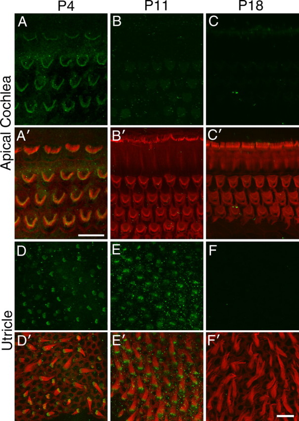
Vlgr1 distribution during postnatal development of the hair bundle. Confocal images of hair bundles from the cochlea (A–C) and utricle (D–F) at P4 (A, D), P11 (B, E), and P18 (C, F) double labeled with anti-Vlgr1 antibodies (green) and Texas Red phalloidin (red). Vlgr1 disappears from hair bundles in the cochlea by P11 and from the utricle by P18. Scale bars: A, 10 μm; F′, 10 μm (applies to B–F).