


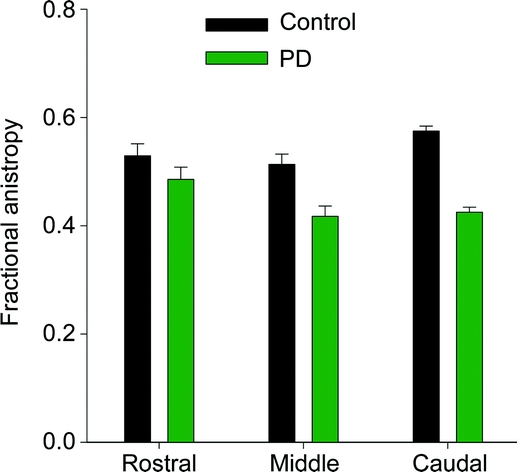
Figure 2 Differences between groups
Mean fractional anisotropy across patients with Parkinson disease (green) and healthy control subjects (black) in the rostral, middle, and caudal region of the substantia nigra. Error bars represent ± 1 SD.
Official websites use .gov
A
.gov website belongs to an official
government organization in the United States.
Secure .gov websites use HTTPS
A lock (
) or https:// means you've safely
connected to the .gov website. Share sensitive
information only on official, secure websites.
Figure 2 Differences between groups
Mean fractional anisotropy across patients with Parkinson disease (green) and healthy control subjects (black) in the rostral, middle, and caudal region of the substantia nigra. Error bars represent ± 1 SD.