



Abstract
Micromolar concentrations of the bile salt deoxycholate are shown to rescue the activity of an inactive mutant, E101A, in the K proton pathway of Rhodobacter sphaeroides cytochrome c oxidase. A crystal structure of the wild-type enzyme reveals, as predicted, deoxycholate bound with its carboxyl group at the entrance of the K path. Since cholate is a known potent inhibitor of bovine oxidase and is seen in a similar position in the bovine structure, the crystallographically defined, conserved steroid binding site could reveal a regulatory site for steroids or structurally related molecules that act on the essential K proton path.
Cytochrome c oxidase (CcO)1 catalyzes a reaction vital to energy production, reducing oxygen to water while translocating protons across the membrane to form the electrochemical gradient used for ATP synthesis. Because of its importance in energy metabolism, a variety of regulatory mechanisms are expected and observed, to achieve control of activity and efficiency. For the mammalian mitochondrial enzyme, besides being controlled on the transcriptional (1), translational (2), and molecular assembly levels (3), its activity is also regulated by secondary messengers such as cAMP (4, 5), Ca2+ (5), and nitric oxide (6), with consequences that are not yet well defined.
The detailed mechanism of how CcO couples electron transfer to proton pumping, and various modes of regulation, are not fully understood. Some mechanistic insight has been gained by studying the bacterial homologues of the mammalian enzyme, such as CcO from Rhodobacter sphaeroides (Rs), by mutational, kinetic, and crystallographic methods. In the bacterial system, it has been established that during each catalytic cycle protons are taken up from the inside of the membrane via two pathways, labeled D and K (7). The D pathway transports protons destined for translocation across the membrane, plus protons used for generation of water at the active site, while the K pathway transports only protons involved in reduction of oxygen to water.
Mutations of a key residue at the entrance of the K path, E101IIA (Rs numbering, with the subscript representing the subunit number), leads to severe inhibition of overall activity (<5% of that of the wild type), but the remaining activity is still coupled to proton pumping. We previously reported that certain carboxylic acids, including arachidonic acid and the bile acid, cholate, can rescue the activity of the E101IIA mutant, from a few percent to more than 50% of the wild-type activity (8). We hypothesize that the rescuing effect is due to the ability of the exogenous lipophilic carboxylate to bind to a position near the entrance of the proton pathway, partially compensating for the loss of the carboxylate group. Arachidonic acid has a similar effect on a D path mutant, D132IA (9), but cholate activates only the K path (8), suggesting a more specific interaction.
Here we report the potent rescuing effect of deoxycholate on the K path mutant of RsCcO (Figure 1), and the X-ray crystal structure of wild-type RsCcO with deoxycholate bound in close proximity to the K pathway, with its carboxyl group positioned at the entrance, as predicted from the rescuing effect (Figure 2).
Figure 1.
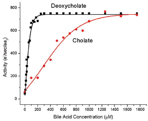
Rescuing effect of cholate (red) and deoxycholate (black) on the activity of E101IIA mutant RsCcO. The steady-state molecular activities of the detergent-solubilized RsCcO were assayed as described in detail in the Supporting Information.
Figure 2.

Deoxycholate resolved near the K path entrance of RsCcO. (A) Overview of the structure of RsCcO containing catalytic core subunits I (green) and II (cyan). The metal centers are shown as spheres (Cu, orange; Fe, red; Mg, blue; Ca, yellow; Cd, wheat). The heme groups are colored by atom type. The two proton uptake channels, D and K paths, are indicated by the black dotted lines. The water molecules within the pathways are shown as red spheres, and some of the key residues are shown as sticks. Note that the resolved deoxycholate molecule (dochl, red sticks) is located near the K path entrance. A detergent molecule, decyl maltoside (DM, blue and red sticks), is also resolved nearby. (B) The 2Fo − Fc difference electron density map (blue) contoured at the 1.0σ level. Residues E101II and H96II are colored by atom type, and Cd is colored yellow (PDB entry 3DTU).
Figure 1 shows the comparison of the activating effects of cholate and deoxycholate on the E101IIA mutant. The final activation level is similar, giving approximately 50% of wild-type activity (700 vs 1200 e−/s), but half-maximal stimulation was achieved at 40 μM for deoxycholate and 350 μM for cholate. This suggests that deoxycholate may bind to RsCcO more tightly and in a more specific manner, which makes it a superior candidate for cocrystallization in determining the liganded structure.
The structure of wild-type CcO with deoxycholate bound was determined at 2.15 Å resolution (Figure 2). A new and more extensive packing interface was present in the deoxycholate-bound versus unbound CcO structure. A deoxycholate molecule (dochl) is clearly resolved, as shown in Figure 2A,B. There are two molecules of CcO in the asymmetric unit, but only one of them has the deoxycholate molecule bound.
Detailed examination of the binding interaction, shown in Figure 3A, reveals that the sterol ring of the deoxycholate is associated with a hydrophobic portion of subunit I that is normally within the lipid bilayer, via multiple van der Waals interactions involving P358I, I361I, P315I, T105II, and I102II. The 3-OH group is hydrogen bonded to the carbonyl oxygen of Y318I and to the side chain of A322I, while the carboxylate group is situated at the K path entrance at the inner surface of the membrane. The binding of the deoxycholate is further strengthened by the ligation of its carboxyl group to a cadmium ion that is also ligated to E101 and H96 of subunit II. Cadmium is a known inhibitor of wild-type RsCcO, as well as an important ingredient for successful crystallization (10). The cadmium ion bound to E101II and H96II is also observed in RsCcO structures without deoxycholate (8, 10).
Figure 3.

Comparison of the conserved steroid binding sites near the K path entrance of CcO. (A) Detailed binding interactions between the deoxycholate and residues in RsCcO (subunit I, green; subunit II, cyan), including those that comprise the K path. Dochl and DM are shown as sticks and colored the same as in Figure 2. Residues that interact with the dochl molecule are shown in sticks and colored by atom type. Cd is shown as a wheat sphere. Part of the hydroxyl farnesyl tail of heme a3 is shown as purple sticks. (B) Comparison of the binding of the steroid molecules between RsCcO and bovine CcO. Subunits I and II of both structures are represented in the same way as in panel A. Subunit VIa from a neighboring CcO molecule in the bovine CcO structure is colored wheat. Dochl resolved in the RsCcO structure is shown as red sticks. The cholate molecule (chl) found in the bovine CcO structure is shown in sticks and colored by atom type (blue and red). Part of the phosphatidylethanolamine molecule (PE) resolved in the bovine CcO structure is shown as yellow sticks. Some residues from both structures that interact with the steroid molecule are shown as sticks. The structure of RsCcO with dochl is labeled in black, while that of bovine CcO with chl is labeled in blue.
In the vicinity of the deoxycholate, a detergent molecule, decyl maltoside (DM), is also clearly resolved (Figures 2B and 3A). This decyl maltoside molecule associates with one side of the deoxycholate, with its alkyl tail forming multiple van der Waals contacts with the hydrophobic edge of the sterol group of deoxycholate, while the polar sugar headgroup forms interactions with the carboxyl group of the deoxycholate. It has been previously shown that certain detergents can mimic the binding of membrane lipids to membrane proteins and occupy their specific binding sites (10). In fact, a lipid in this position, also associated with the hydrophobic surface of cholate, is resolved in the bovine CcO structure (Figure 3B). Therefore, it is conceivable that in the native membrane, a lipid is present at this site in RsCcO as well and could contribute to the stabilization of steroid binding.
It has long been known that cholate, while being widely used as a solubilizing agent in preparing bovine mitochondrial CcO, promotes dimerization of the enzyme (11) and is also a strong reversible inhibitor (12). The mechanism for such inhibition is unclear, but electron transfer between heme a and heme a3 is observed to be markedly slowed in the presence of cholate (13). In the structure of bovine CcO (PDB entry 2DYR, 1.8Å), which is a homodimer, a total of four cholate molecules are resolved per monomer, two of them at the dimer interface (14). These cholate molecules bind to residues in both monomers of the dimer, but for the site that matches the RsCcO binding site (Figure 3B), the interaction with a nuclear subunit, VIa, was considered to be of primary interest (15). Careful examination of this bovine CcO VIa binding site shows that the cholate is also bound to subunits I and II of the other monomer, near the entrance of the K pathway. The position is similar to and involves some of the same residues as the deoxycholate bound in RsCcO (Figure 3B). The binding of cholate at this position in bovine CcO is different in detail from the deoxycholate binding in Rs, which could account for the observed inhibitory effect in bovine, not seen in wild-type RsCcO (8). In bovine CcO, the carboxyl oxygen of E62II at the K pathway entrance (the equivalent of E101II in Rs) is tightly hydrogen bonded to the 7-OH group of cholate, possibly affecting proton transport.
It was previously proposed that the binding sites of cholate in the bovine CcO structure indicated potential nucleotide binding sites, since cholate is similar to ADP in size and shape (16). The two proposed nucleotide binding sites are at the interface of the dimer: one between subunits I and III close to a conserved Y304 (Y347 in RsCcO) of subunit I which is phosphorylated in bovine CcO (4) and the other in the same location as the RsCcO deoxycholate site and also close to a known site of phosphorylation in bovine CcO, T11 of subunit VIa (14). There is considerable evidence for cholate and adenine nucleotide regulation of the mammalian CcO (15).
Given the previous observations of regulation of CcO activity by a variety of ligands, including bile salts (12) and glucocorticoids (17), and our current evidence of a conserved site that impacts an essential proton uptake path, it is reasonable to propose that the conserved site identified in this study could represent a physiological binding site for steroids or other effector molecules. This possibility is being explored.
Acknowledgments
We thank Dr. R. Michael Garavito from the Biochemistry and Molecular Biology Department, Michigan State University, for his support and consultation, and beamline staff scientists at LS-CAT and GM/CA-CAT, Advanced Photon Source, Argonne National Laboratory, Argonne, IL, for their help during data collection.
Footnotes
Supported by National Institutes of Health Grant GM26916, Michigan Technology Tri Corridor Center for Structural Biology Core Technology Alliance Grant 085P1000817, and Michigan State University Research Excellence Fund Grant 03-016.
Structure files deposited as Protein Data Bank entry 3DTU.
Abbreviations: CcO, cytochrome c oxidase; Rs, Rhodobacter sphaeroides; DM, decyl maltoside; dochl, deoxycholate; chl, cholate; PDB, Protein Data Bank.
SUPPORTING INFORMATION AVAILABLE
Details of protein purification, oxidase activity assays, crystallization, crystallographic data collection, and structure refinement. This material is available free of charge via the Internet at http://pubs.acs.org.
References
- 1.Lenka N, Vijayasarathy C, Mullick J, Avadhani NG. Prog Nucleic Acid Res Mol Biol. 1998;61:309–344. doi: 10.1016/s0079-6603(08)60830-2. [DOI] [PubMed] [Google Scholar]
- 2.Aschrafi A, Schwechter A, Natera O, Gioio A, Kaplan B. J Neurochem. 2008;104(1):34. [Google Scholar]
- 3.Fontanesi F, Soto Ileana C, Horn D, Barrientos A. Am J Physiol. 2006;291:C1129–C1147. doi: 10.1152/ajpcell.00233.2006. [DOI] [PubMed] [Google Scholar]
- 4.Lee I, Salomon AR, Ficarro S, Mathes I, Lottspeich F, Grossman LI, Hüttemann M. J Biol Chem. 2005;280:6094–6100. doi: 10.1074/jbc.M411335200. [DOI] [PubMed] [Google Scholar]
- 5.Ludwig B, Bender E, Arnold S, Hüttemann M, Lee I, Kadenbach B. ChemBioChem. 2001;2:392–403. doi: 10.1002/1439-7633(20010601)2:6<392::AID-CBIC392>3.0.CO;2-N. [DOI] [PubMed] [Google Scholar]
- 6.Brunori M, Giuffre A, Forte E, Mastronicola D, Barone MC, Sarti P. Biochim Biophys Acta. 2004;1655:365–371. doi: 10.1016/j.bbabio.2003.06.008. [DOI] [PubMed] [Google Scholar]
- 7.Svensson-Ek M, Abramson J, Larsson G, Tornroth S, Brzezinski P, Iwata S. J Mol Biol. 2002;321:329–339. doi: 10.1016/s0022-2836(02)00619-8. [DOI] [PubMed] [Google Scholar]
- 8.Qin L, Mills DA, Hiser C, Murphree A, Garavito RM, Ferguson-Miller S, Hosler J. Biochemistry. 2007;46:6239–6248. doi: 10.1021/bi700173w. [DOI] [PMC free article] [PubMed] [Google Scholar]
- 9.Fetter J, Sharpe M, Qian J, Mills D, Ferguson-Miller S, Nicholls P. FEBS Lett. 1996;393:155–160. doi: 10.1016/0014-5793(96)00874-5. [DOI] [PubMed] [Google Scholar]
- 10.Qin L, Hiser C, Mulichak A, Garavito RM, Ferguson-Miller S. Proc Natl Acad Sci USA. 2006;103:16117–16122. doi: 10.1073/pnas.0606149103. [DOI] [PMC free article] [PubMed] [Google Scholar]
- 11.Musatov A, Robinson NC. Biochemistry. 2002;41:4371–4376. doi: 10.1021/bi016080g. [DOI] [PubMed] [Google Scholar]
- 12.Van Buuren KJH, Van Gelder BF. Biochim Biophys Acta. 1974;333:209–217. doi: 10.1016/0005-2728(74)90005-x. [DOI] [PubMed] [Google Scholar]
- 13.Yu C-A, Lu L, King TE. J Biol Chem. 1975;250:1383–1392. [PubMed] [Google Scholar]
- 14.Shinzawa-Itoh K, Aoyama H, Muramoto K, Terada H, Kurauchi T, Tadehara Y, Yamasaki A, Sugimura T, Kurono S, Tsujimoto K, Mizushima T, Yamashita E, Tsukihara T, Yoshikawa S. EMBO J. 2007;26:1713–1725. doi: 10.1038/sj.emboj.7601618. [DOI] [PMC free article] [PubMed] [Google Scholar]
- 15.Kadenbach B, Napiwotzki J, Frank V, Arnold S, Exner S, Hüttemann M. J Bioenerg Biomembr. 1998;30:25–33. doi: 10.1023/a:1020599209468. [DOI] [PubMed] [Google Scholar]
- 16.Tsukihara T, Aoyama H, Yamashita E, Tomizaki T, Yamaguchi H, Shinzawa-Itoh K, Nakashima R, Yaono R, Yoshikawa S. Science. 1996;272:1136–1144. doi: 10.1126/science.272.5265.1136. [DOI] [PubMed] [Google Scholar]
- 17.Simon N, Jolliet P, Morin C, Zini R, Urien S, Tillement J-P. FEBS Lett. 1998;435:25–28. doi: 10.1016/s0014-5793(98)01033-3. [DOI] [PubMed] [Google Scholar]