


Fig. 2.
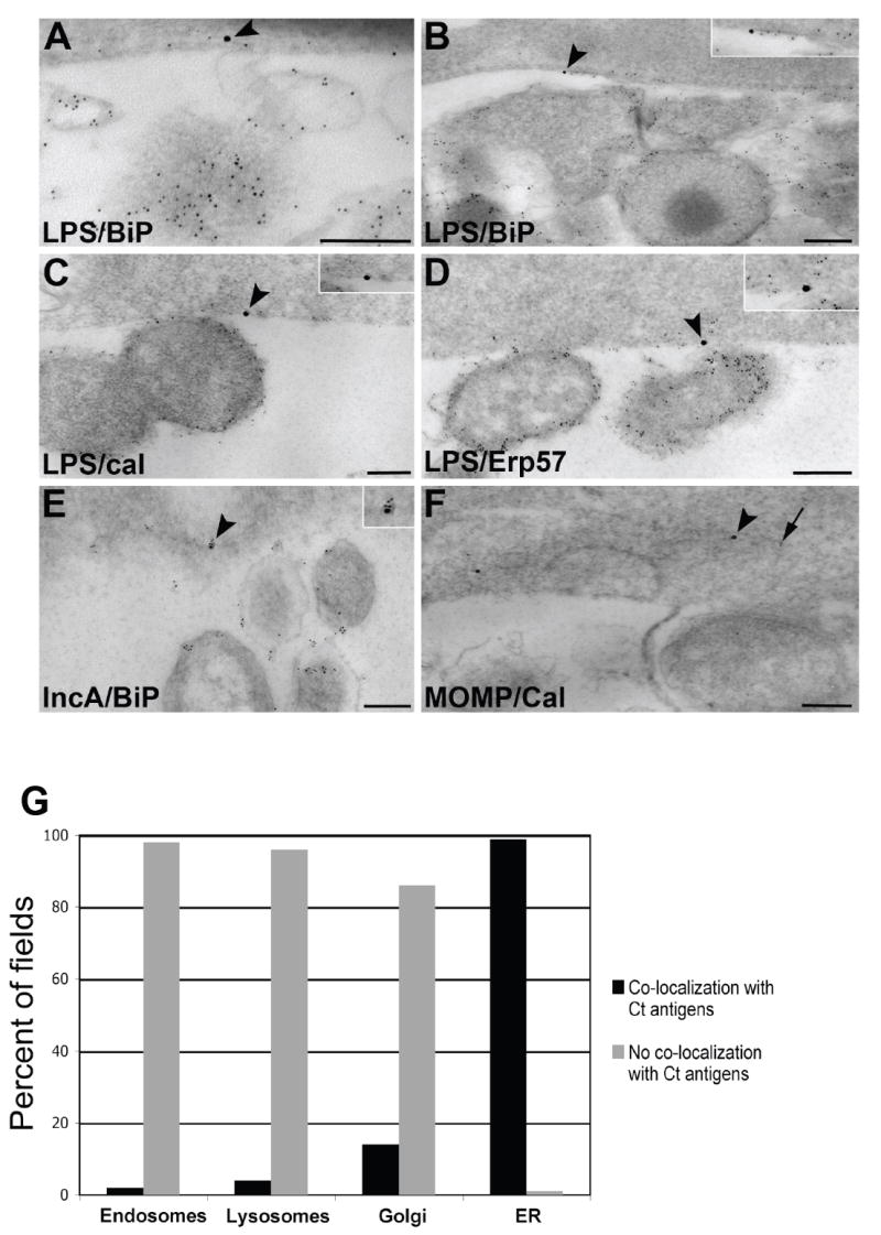
Ultrastructural co-localization between chlamydial antigens and host cell ER markers. HEC-1B cells infected with C. trachomatis E for 48 h were prepared for post-embedding immunoelectron microscopy. (A-D) Co-localization between chlamydial LPS (5 nm gold; arrow) and the ER markers BiP/GRP78 (A and B), calnexin (C) and Erp57 (D; 15 nm gold; arrowheads; magnified in insets). Scale bars, 200 nm. (E) Co-localization between chlamydial IncA (5 nm gold) and the ER marker BiP/GRP78 (15 nm gold; arrowhead, magnified in inset). Scale bar, 200 nm. (F) Co-localization between chlamydial MOMP (5 nm gold; arrow) and the ER marker calnexin (15 nm gold; arrowhead). Scale bar, 200 nm. (G) Quantitation of the co-localization between chlamydial antigens and host cell organelles. Multiple images (>50 fields per host cell organelle) were examined and percentages calculated for graphical representation. Levels of significance (all p-values<0.001) indicate co-localization of chlamydial antigens with the ER, but not with endosomes, lysosomes nor Golgi.