


Fig 3.
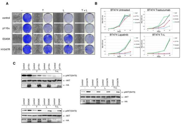
Constitutive activation of the PI3K pathway decreases sensitivity to trastuzumab and lapatinib. When required cells were either treated with trastuzumab (5 ug/ml), or lapatinib (27 nM) unless otherwise stated. A) Colony formation assay in BT474 cells infected with either WT PIK3CAα or PIK3CAα mutants E545K and H1047R, or relevant controls. Infected cells were treated with trastuzumab, or lapatinib, or both. After 4 weeks cells were photographed and stained with crystal violet. B) Growth curves of stably infected PI3K wildtype or mutant PIK3CAα BT474 cells treated for 3 weeks with trastuzumab, or lapatinib, or both. Cell numbers were quantified as described in Experimental Procedures. Growth curves were performed in triplicate with the error bars depicting the mean ± s.d. C) Western blot analysis of BT474 cells infected with HA-tagged PI3K PIK3CAα (upper left panel), PI3K(E545K) PIK3CAα (lower left panel), PI3K(H1047R) PIK3CAα (right panel), or controls were treated overnight with trastuzumab, or lapatinib, or both. Whole cell extracts were analyzed with indicated antibodies.